

Nanoparticle approaches for the renin-angiotensin system
- PMID: 37484281
- PMCID: PMC10361043
- DOI: 10.1016/j.heliyon.2023.e16951
Nanoparticle approaches for the renin-angiotensin system
Abstract
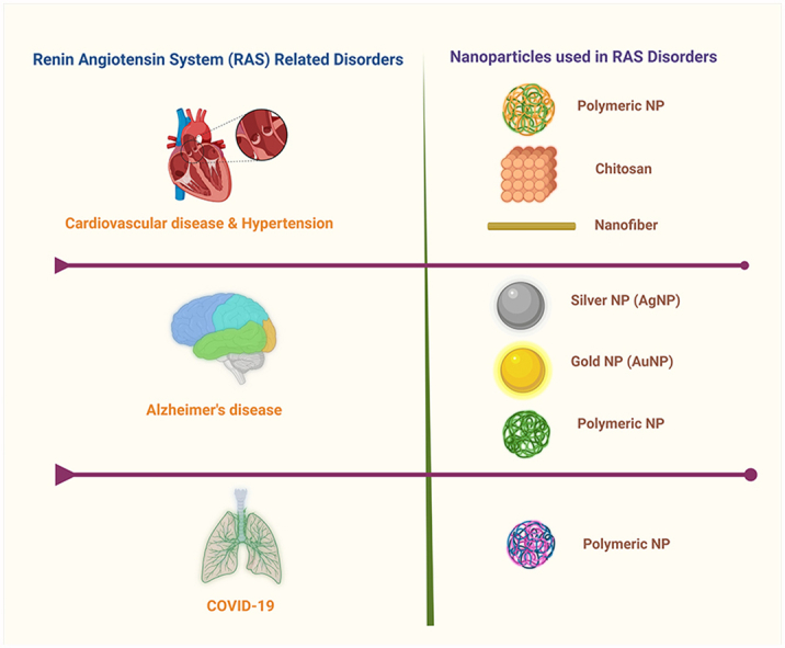
The renin-angiotensin system (RAS) is a hormonal cascade that contributes to several disorders: systemic hypertension, heart failure, kidney disease, and neurodegenerative disease. Activation of the RAS can promote inflammation and fibrosis. Drugs that target the RAS can be classified into 3 categories, AT1 angiotensin receptor blockers (ARBs), angiotensin-converting enzyme (ACE) inhibitors, and renin inhibitors. The therapeutic efficacy of current RAS-inhibiting drugs is limited by poor penetration across the blood-brain barrier, low bioavailability, and to some extent, short half-lives. Nanoparticle-mediated drug delivery systems (DDSs) are possible emerging alternatives to overcome such limitations. Nanoparticles are ideally 1-100 nm in size and are considered efficient DDSs mainly due to their unique characteristics, including water dispersity, prolonged half-life in blood circulation, smaller size, and biocompatibility. Nano-scale DDSs can reduce the drug dosage frequency and acute toxicity of drugs while enhancing therapeutic success. Different types of nanoparticles, such as chitosan, polymeric, and nanofibers, have been examined in RAS-related studies, especially in hypertension, cardiovascular disease, and COVID-19. In this review article, we summarize the physical and chemical characteristics of each nanoparticle to elaborate on their potential use in RAS-related nano-drug delivery research and clinical application.
Keywords: COVID-19; Cardiovascular disease; Drug delivery; Hypertension; Nanoparticles; Renin-angiotensin system.
© 2023 Published by Elsevier Ltd.
Conflict of interest statement
The authors declare the following financial interests/personal relationships which may be considered as potential competing interests: Robert C. Speth reports financial support was provided by National Institutes of Health. The authors declare no conflict of interest.
Figures


References
-
- Tigerstedt R., Bergmann P.G. Niere und kreislauf. Scand. Arch. Physiol. 1898;8:223–271.
-
- Speth R.C. Reference Module in Biomedical Sciences. Elsevier; 2021. Renin-angiotensin-aldosterone system.
-
- Rodrigues Prestes T.R., Rocha N.P., Miranda A.S., Teixeira A.L., Simoes-e-Silva A.C. The anti-inflammatory potential of ACE2/angiotensin-(1-7)/Mas receptor axis: evidence from basic and clinical research. Curr. Drug Targets. 2017;18:1301–1313. - PubMed
Publication types
LinkOut - more resources
Full Text Sources
Research Materials
Miscellaneous
