

Carotid body paraganglioma: a case report
- PMID: 37484598
- PMCID: PMC10362654
- DOI: 10.11604/pamj.2023.44.182.38636
Carotid body paraganglioma: a case report
Abstract
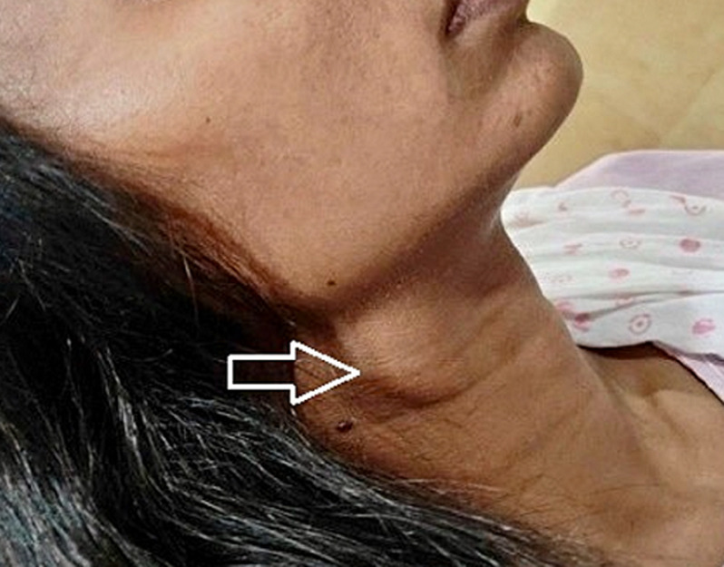
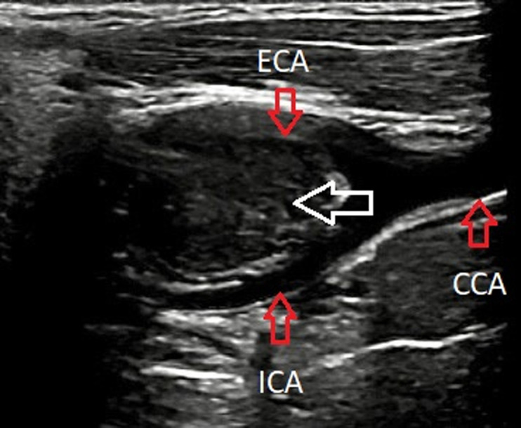
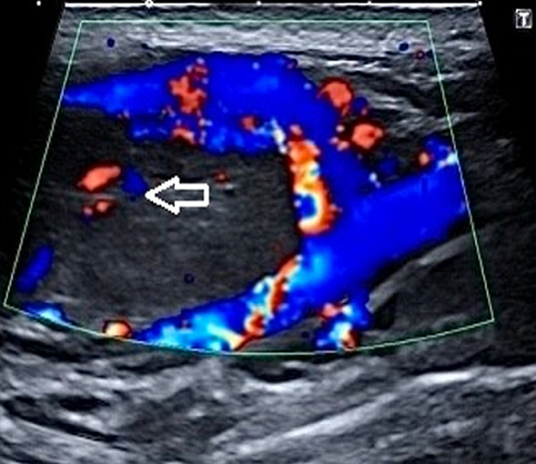
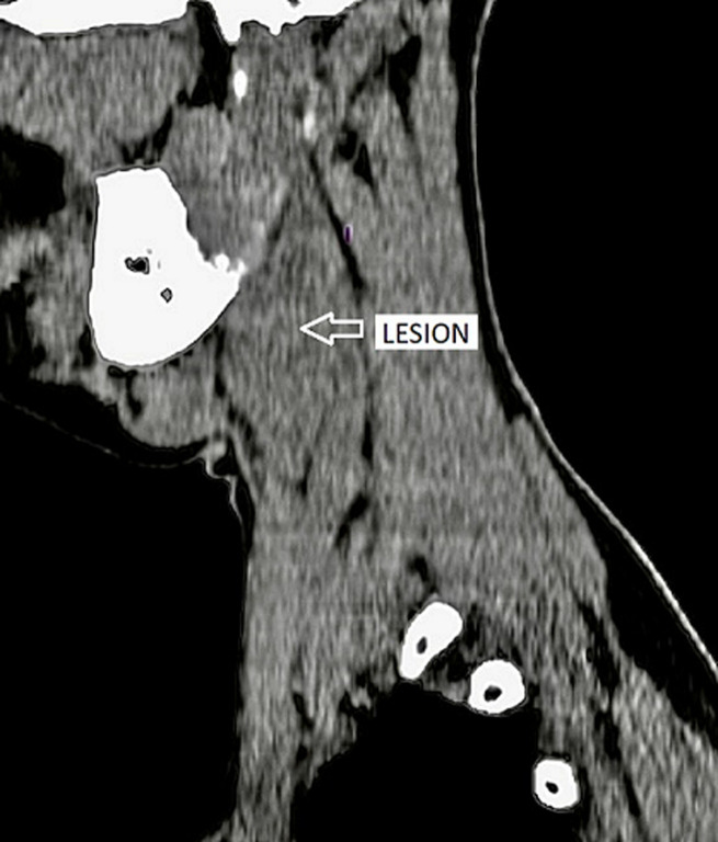
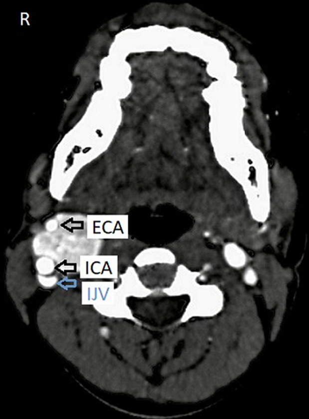
A highly vascular glomus tumor that develops from the paraganglion cells of the carotid body is called a carotid body tumor (CBT), also known as a chemodectoma or carotid body paraganglioma (CBP). It is situated near the carotid bifurcation, where the external and internal carotid arteries splay out characteristically. We present a case of a 30-year-old woman who had a slightly tender, slightly pulsatile, and slightly ballotable swelling over the lateral aspect of the neck on the right side. The surgical resection of the tumor was done based on the diagnosis made on clinical-radiological investigations as a carotid body tumor further confirmed by a histopathological study. We also provide a summary of the research on carotid body tumors clinical and imaging manifestations, assessment, and therapy.
Keywords: Paraganglioma; carotid body tumor; case report; chemodectomas; neck tumor.
Copyright: Swaragandha Shivaji Jadhav et al.
Conflict of interest statement
The authors declare no competing interests.
Figures

References
-
- García MA, Pendás JL, Tapia JP, Rostán GG, Fente VS, Pelaz AC, et al. Head and neck paragangliomas: revision of 89 cases in 73 patients. Acta Otorrinolaringol Esp. 2007 Mar;58(3):94–100. - PubMed
-
- Duncan AW, Lack EE, Deck MF. Radiological evaluation of paragangliomas of the head and neck. Radiology. 1979 Jul;132(1):99–105. - PubMed
-
- Lattes R. Nonchromaffin paraganglioma of ganglion nodosum, carotid body, and aortic-arch bodies. Cancer. 1950 Jul;3(4):667–94. - PubMed
Publication types
MeSH terms
LinkOut - more resources
Full Text Sources
