Review
. 2023 Jun 3;8(2):36-48.
doi: 10.22575/interventionalradiology.2022-0008. eCollection 2023 Jul 1.

Endovascular and Percutaneous Embolotherapy for the Body and Extremity Arteriovenous Malformations

Affiliations
Review

Endovascular and Percutaneous Embolotherapy for the Body and Extremity Arteriovenous Malformations

Keigo Osuga et al. Interv Radiol (Higashimatsuyama). .

Abstract

Arteriovenous malformations (AVMs) consist of abnormal communications between the arteries and veins. They can involve any part of the body and extremity and grow in proportion to age and in response to hormonal influence or trauma. When symptoms progress from Schöbinger clinical stage II to III, transcatheter and/or direct puncture embolization are less-invasive and repeatable options for symptom palliation. The goal of embolization is to obliterate the AV shunt, and the choice of lesion access and embolic agents is based on the individual anatomy and flow. Embolization can be technically challenging due to complex vascular anatomy and morbidity risks. Therefore, a multidisciplinary management is essential for the diagnosis and therapeutic intervention of AVMs.

Keywords: arteriovenous fistula; arteriovenous malformations; arteriovenous shunt; embolic agents; embolization.


Conflict of interest statement

None

Figures

Figure 1.
Figure 1.
A female in her 50s with Cho’s type-I AVM of the left buttock. She suffered from left-buttock pain (Reprinted from Ref. . with permission). a The left internal iliac arteriogram shows a direct AVF between the distal branch of the internal iliac artery and an aneurysmal dominant outflow vein (DOV) (arrow). b A 5Fr balloon catheter was placed in the internal iliac artery for arterial inflow control. A microcatheter reached the DOV, and detachable microcoils were deployed as scaffold. 0.7 mL of 20% NBCA-lipiodol mixture was injected into the space among the framing coils in DOV back to the end of the feeding artery. c Final angiography shows disappearance of the AV shunt.
Figure 2.
Figure 2.
A female in her 40s with Cho’s type-IIIa AVM of the left foot. a The left popliteal arteriogram shows micro-AV shunts both in the dorsal and planter aspects of the foot (arrows). Stepwise direct puncture embolization was performed three times at 4 months intervals. The AV shunt was punctured by 25G needles, and 0.5–1.0 mL of absolute ethanol was injected at 5–10-min intervals under tourniquet control. b Post-embolization angiography at the third session shows nearly total disappearance of the AV shunt. c A photograph before embolization. The patient suffered from intractable pain and skin ulcer. d A photograph 3 years after the last embolization. The skin ulcer was gradually healed.
Figure 3.
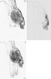
Figure 3.
A male in his 60s with Yakes type-IV AVM of the left hand. He suffered from pain, swelling, and deformity of the palm. a The left radial arteriogram shows diffuse capillary-like stain in the palm and the thumb. Transarterial embolization was performed from each feeding artery using a 1.5Fr flow-guided microcatheter. A total of 7 mL of 50% diluted ethanol was intermittently injected. b Selective arteriogram of the feeding artery into the thumb shows micro-AV shunts. c Post-embolization angiography shows mild decrease in the shunt with appearance of the digital arteries in the second and third fingers.

References

    1. Tasiou A, Tzerefos C, Alleyne CH Jr, et al. Arteriovenous malformations: congenital or acquired lesions? World Neurosurg. 2020; 134: e799-e807 - PubMed
    1. Greene AK, Liu AS, Mulliken JB, Chalache K, Fishman SJ. Vascular anomalies in 5621 patients: guidelines for referral. J Pediatr Surg. 2011; 46: 1784-1789. - PubMed
    1. Kohout MP, Hansen M, Pribaz JJ, Mulliken JB. Arteriovenous malformations of the head and neck: natural history and management. Plast Reconstr Surg. 1998; 102: 643-654. - PubMed
    1. Liu AS, Mulliken JB, Zurakowski D, Fishman SJ, Greene AK. Extracranial arteriovenous malformations: natural progression and recurrence after treatment. Plast Reconstr Surg. 2010; 125: 1185-1194. - PubMed
    1. Mimura H, Akita S, Fujino A, et al. Japanese clinical practice guidelines for vascular anomalies 2017. Jpn J Radiol. 2020; 38: 287-342. - PMC - PubMed