

Peristomal adenocarcinoma 16 years after colorectal adenocarcinoma resection with curative intent
- PMID: 37485497
- PMCID: PMC10359180
- DOI: 10.1093/jscr/rjad419
Peristomal adenocarcinoma 16 years after colorectal adenocarcinoma resection with curative intent
Abstract
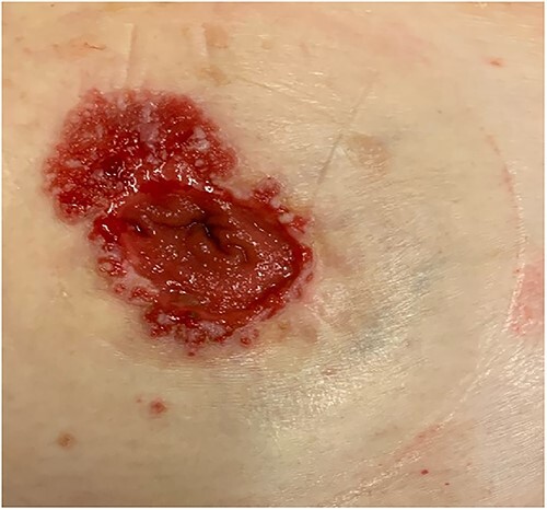
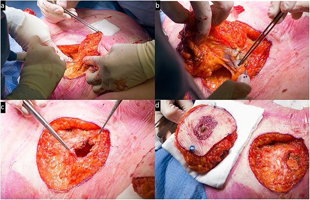
Metachronous colorectal cancer is relatively rare, occurring in 0.7-3.6% of patients diagnosed with colorectal adenocarcinoma. Cutaneous metastases are similarly a rare presentation, occurring in <6% of metastatic colorectal cancer patients. Even more rare are the cutaneous recurrences at the peristomal site. Clinically, it is difficult to distinguish between metachronous cancer and cutaneous metastases. This paper reports a case of an elderly woman presenting with a slowly progressing peristomal cutaneous lesion 16 years after surgical resection for colorectal cancer. Core punch biopsy revealed a cutaneous localization of an intestinal type of adenocarcinoma. A surgical resection of the peristomal area was carried out whereby a new colostomy was created on the contralateral side. Definite histopathological examination showed a superficially located intestinal type adenocarcinoma with extensive pagetoid spread in the epidermal surface. In conclusion, it is important to remain alert and strive for early detection for cutaneous abnormalities following colorectal cancer.
Published by Oxford University Press and JSCR Publishing Ltd. © The Author(s) 2023.
Conflict of interest statement
None declared.
Figures


References
-
- Federatie Medisch Specialisten . Colorectaal carcinoom (CRC): Follow-up na chirurgische resectie stadium I-III colon- en rectumcarcinoom. Richtlijnendatabase 2020.
-
- Park I, Yu C, Kim H, Jung Y, Han K, Kim J. Metachronous colorectal cancer. Colorectal Dis 2006;8:323–7. - PubMed
-
- Kuo Y-H, Chin C-C, Lee K-F. Metastasis at the colostomy site: a rare case report. Jpn J Clin Oncol 2012;42:753–6. - PubMed
Publication types
LinkOut - more resources
Full Text Sources
