. 2023 Aug 31;14(4):e0148223.
doi: 10.1128/mbio.01482-23. Epub 2023 Jul 24.

CD9 co-operation with syndecan-1 is required for a major staphylococcal adhesion pathway

Affiliations

CD9 co-operation with syndecan-1 is required for a major staphylococcal adhesion pathway

Luke R Green et al. mBio. .

Abstract

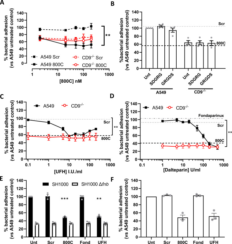
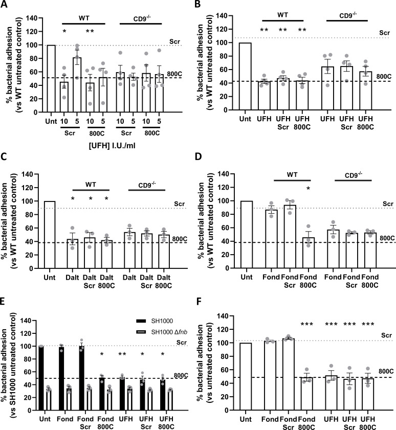
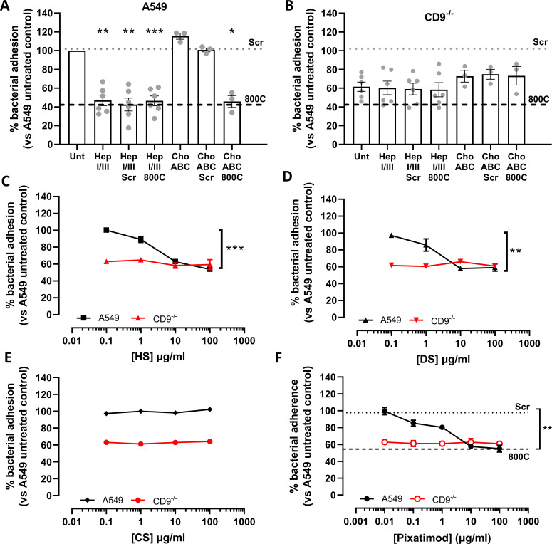
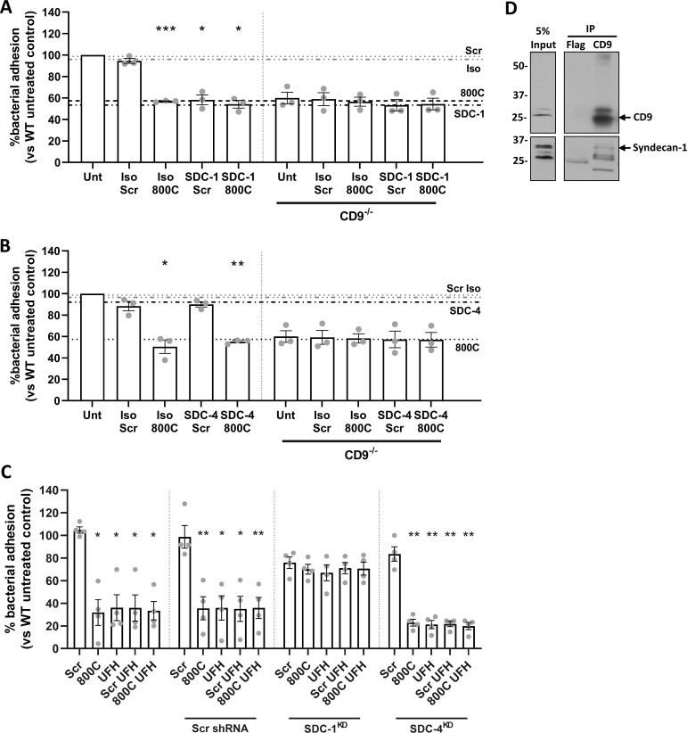
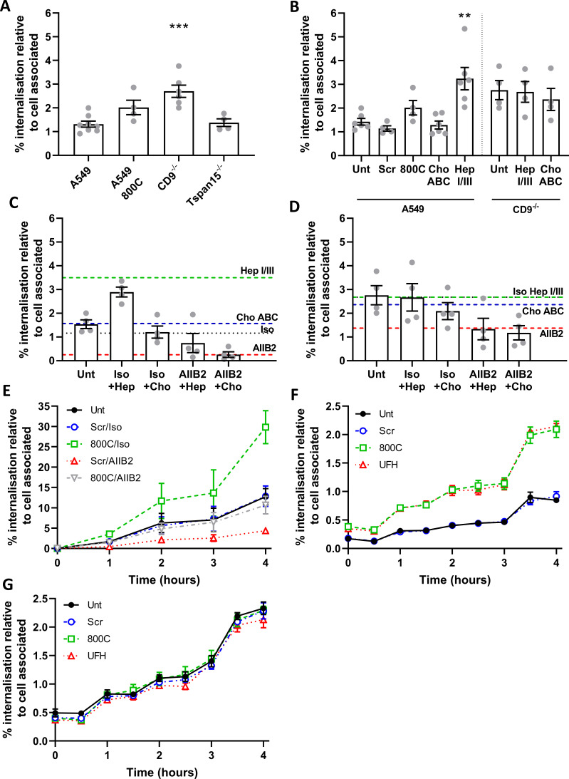
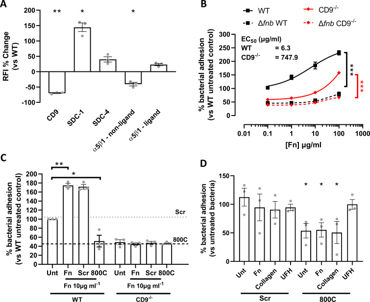
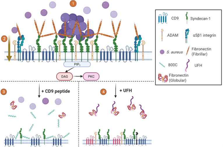
Epithelial colonization is a critical first step in bacterial pathogenesis. Staphylococcus aureus can utilize several host factors to associate with cells, including α5β1 integrin and heparan sulfate proteoglycans, such as the syndecans. Here, we demonstrate that a partner protein of both integrins and syndecans, the host membrane adapter protein tetraspanin CD9, is essential for syndecan-mediated staphylococcal adhesion. Fibronectin is also essential in this process, while integrins are only critical for post-adhesion entry into human epithelial cells. Treatment of epithelial cells with CD9-derived peptide or heparin caused significant reductions in staphylococcal adherence, dependent on both CD9 and syndecan-1. Exogenous fibronectin caused a CD9-dependent increase in staphylococcal adhesion, whereas blockade of β1 integrins did not affect adhesion but did reduce the subsequent internalization of adhered bacteria. CD9 disruption or deletion increased β1 integrin-mediated internalization, suggesting that CD9 coordinates sequential staphylococcal adhesion and internalization. CD9 controls staphylococcal adhesion through syndecan-1, using a mechanism that likely requires CD9-mediated syndecan organization to correctly display fibronectin at the host cell surface. We propose that CD9-derived peptides or heparin analogs could be developed as anti-adhesion treatments to inhibit the initial stages of staphylococcal pathogenesis. IMPORTANCE Staphylococcus aureus infection is a significant cause of disease and morbidity. Staphylococci utilize multiple adhesion pathways to associate with epithelial cells, including interactions with proteoglycans or β1 integrins through a fibronectin bridge. Interference with another host protein, tetraspanin CD9, halves staphylococcal adherence to epithelial cells, although CD9 does not interact directly with bacteria. Here, we define the role of CD9 in staphylococcal adherence and uptake, observing that CD9 coordinates syndecan-1, fibronectin, and β1 integrins to allow efficient staphylococcal infection. Two treatments that disrupt this action are effective and may provide an alternative to antibiotics. We provide insights into the mechanisms that underlie staphylococcal infection of host cells, linking two known adhesion pathways together through CD9 for the first time.

Keywords: CD9; HSPG; Staphylococcus aureus; bacterial adhesion; epithelial; fibronectin; syndecan; tetraspanin.


Conflict of interest statement

P.N.M., R.I., and B.C. are co-inventors on patent WO2021175809A1, related to the peptides used in this study. All other authors declare that the research was conducted in the absence of any commercial or financial relationships that could be construed as a potential conflict of interest.

Figures

Fig 1
Fig 1
Tetraspanin derived peptides and heparin derivatives but not RGD peptides reduce staphylococcal adherence. Cells were infected with SH1000 for 60 min at an MOI = 50. (A) WT (black) or CD9−/− (red) cells treated with scrambled (dotted) or 800C peptide (solid) for 60 min prior to infection. WT (black) or CD9−/− (red) cells treated with RGD peptides (100 µM) (B), or various concentrations of heparin sodium (C) or dalteparin (D) for 60 min prior to infection. Effect of CD9-derived peptide treatment (200 nM) shown by dotted lines (B−D). Effect of fondaparinux (10 µg/mL) treatment on WT cells shown in panel D. (E) WT cells treated with scrambled peptide, 800C, fondaparinux, or UFH were infected with either SH1000 (black bars) or SH1000 Δfnb (white bars) for 60 min at an MOI = 50. (F) NTKs treated with scrambled peptide, 800C, fondaparinux, or UFH were infected with SH1000 for 60 min at an MOI = 50. n ≥ 3, mean ± SEM, one-way ANOVA.
Fig 2
Fig 2
Combination treatment of unfractionated heparin and 800C produces no additive effects. Cells were infected with SH1000 for 60 min at an MOI = 50. (A) WT or CD9−/− A549 cells were treated with UFH, 800C peptide, or a combination of both for 60 min prior to treatment. (B) WT or CD9-/- A549 cells were treated with peptide (200 nM) for 60 mins prior to infection. UFH (10 U/ml), either in combination with 800C peptide or as a singular treatment, was added at the start of infection. (D) WT or CD9−/− cells were treated with fondaparinux (10 µg/mL), 800C peptide (200 nM), or a combination of both 60 min prior to infection. (E) WT cells treated with 800C peptide (200 nM), UFH (10 U/mL), or a combination of the two and infected with either SH1000 or SH1000 Δfnb at an MOI = 50. (F) NTKs treated with 800C peptide (200 nM), UFH (10 I.U./mL) or a combination of the two and infected with SH1000 for 60 min at an MOI = 50. n ≥ 3, mean ± SEM, and one-way ANOVA.
Fig 3
Fig 3
Heparan sulfates are required during tetraspanin-mediated staphylococcal adherence. Cells were infected with SH1000 for 60 min at an MOI = 50. (A) WT cells were treated with either 0.25 U/mL chondroitinase ABC or 0.5 U/mL heparinase I/III for 3 h prior to infection. Peptide was added to cells 60 min prior to infection for combination treatments. (B) CD9−/− cells were treated with either 0.25 U/mL chondroitinase ABC or 0.5 U/mL heparinase I/III for 3 h prior to infection. Peptide was added to cells 60 min prior to infection for combination treatments. WT or CD9−/− cells were treated with heparan sulfate (HS; C), dermatan sulfate (DS; D), chondroitin sulfate (CS; E), or the heparan sulfate mimetic (pixatimod; F) for 60 min prior to infection. n ≥ 3, mean ± SEM, and one-way ANOVA.
Fig 4
Fig 4
Syndecan-1 is involved in tetraspanin-mediated staphylococcal adherence to epithelial cells. Cells were infected for 60 min with SH1000 at an MOI = 50. (A) WT or CD9−/− cells were treated with peptides (200 nM), isotype control (JC1), anti-SDC-1 antibodies (B-A38, 10 µg/mL), or a combination of peptide and antibodies for 60 min prior to infection. (B) WT or CD9−/− cells were treated with peptides (200 nM), isotype control (02–6200), anti-SDC-4 antibodies (5G9, 20 µg/mL), or a combination of peptide and antibodies for 60 min prior to infection. (C) shRNA knockdowns of SDC-1 and SDC-4 were treated with peptides (200 nM), UFH (10 I.U./mL), or a combination of the two for 60 min prior to infection. Scrambled shRNA was used as a control. n ≥ 3, mean ± SEM, and one-way ANOVA. (D) Whole cell lysates were immunoprecipitated with anti-CD9 antibodies (MM2/57), and the resulting elutes were probed with either anti-CD9 or anti-SDC-1 antibodies. Lysates were probed with anti-FLAG antibodies as a control.
Fig 5
Fig 5
Interference with CD9 or heparan sulfates increases staphylococcal internalization, which is controlled by α5β1. Cells were infected with SH1000 for 60 min at an MOI = 50. After infection, cells were washed and treated with 200 µg/mL gentamicin sulfate to remove adherent bacteria and lysed to quantify the internalized bacteria. Internalized bacteria were normalized against the cell associated bacteria. (A) Internalized bacteria within WT, CD9−/− , and Tspan15−/− cells, 800C treated WT cells are added as an example. (B) WT or CD9−/− cells were treated with 0.25 U/mL chondroitinase ABC or 0.5 U/mL heparinase I/III for 3 h prior to infection. WT (C) or CD9−/− (D) cells were treated with heparinase I/III or chondroitinase ABC for 3 h. Cells were treated with isotype control or anti-α5β1 antibodies (AIIB2) for 60 min prior to infection. (E) Cells were treated with combinations of peptide (200 nM), isotype control, and anti-α5β1 antibodies (AIIB2) for 60 min prior to infection. Cells were infected for 60 min at 4°C, after infection cells were warmed to 37°C and internalization was allowed to continue for 4 h. WT (F) or CD9−/− (G) cells were treated with scrambled peptide (blue) (200 nM), 800C (green) (200 nM), or UFH (red) (10 I.U./mL) for 60 min prior to infection. Cells were infected for 60 min before being washed and treated with 200 µg/mL gentamicin sulfate. Internalized bacteria were allowed to grow for a further 4 hours before cells were lysed and enumerated. n ≥ 3, mean ± SEM, and one-way ANOVA.
Fig 6
Fig 6
Addition of exogenous Fn increases staphylococcal adherence, which is negated in the presence of 800C. (A) Cell surface expression of receptors of interest was determined in CD9−/− cells. Relative fluorescence intensity was calculated by dividing the test antibody by the isotype control. Percentage change was calculated against WT cell values. (B) Cells were treated with varying concentrations of Fn prior to infection with SH1000 or SH1000 Δfnb. (C) Fn was added to WT or CD9−/− cells in combination with 800C or a scrambled peptide for 1 h prior to infection. (D) SH1000 was pre-treated with Fn, collagen, or UFH for 90 min prior to infection. Cells were treated with 200 nM 800C or scrambled peptide for 60 min prior to infection. n ≥ 3, mean ± SEM, and one-way ANOVA.
Fig 7
Fig 7
Proposed mechanism for tetraspanin-mediated staphylococcal adherence. (1) CD9 enriched microdomains contain syndecan-1. Clustering of syndecan-1 by CD9 recruits fibronectin and produces “adhesion nets”. S. aureus utilizes these nets to initiate initial adherence to the cell surface (2). Inclusion of integrins in CD9 enriched microdomains means the “adhesion nets” are in close proximity to β1 integrins, allowing for the transfer of bacteria after initial adherence for rapid internalization through the canonical pathway. Interactions with CD9 and ADAMs ensure integrins are kept in an inactive state inhibiting bacterial internalization (3). CD9-derived peptides increase the area of TEMs pushing syndecan clusters apart and reducing the formation of “adhesion nets” (4). UFH treatment displaces fibronectin or blocks staphylococcal interaction with Fn, destabilizing TEMs and reducing bacterial adhesion. Mechanisms other than CD9-mediated adhesion and internalization are still possible with both treatments. Created with Biorender.com.

References

    1. Tong SYC, Davis JS, Eichenberger E, Holland TL, Fowler VG. 2015. Staphylococcus aureus infections: epidemiology, pathophysiology, clinical manifestations, and management. Clin Microbiol Rev 28:603–661. doi:10.1128/CMR.00134-14 - DOI - PMC - PubMed
    1. Stryjewski ME, Corey GR. 2014. Methicillin-resistant Staphylococcus aureus: an evolving pathogen. Clin Infect Dis 58:S10–9. doi:10.1093/cid/cit613 - DOI - PubMed
    1. Josse J, Laurent F, Diot A. 2017. Staphylococcal adhesion and host cell invasion: fibronectin-binding and other mechanisms. Front Microbiol 8:2433. doi:10.3389/fmicb.2017.02433 - DOI - PMC - PubMed
    1. Sinha B, François PP, Nüsse O, Foti M, Hartford OM, Vaudaux P, Foster TJ, Lew DP, Herrmann M, Krause KH. 1999. Fibronectin-binding protein acts as Staphylococcus aureus invasin via fibronectin bridging to integrin α5β1. Cell Microbiol 1:101–117. doi:10.1046/j.1462-5822.1999.00011.x - DOI - PubMed
    1. Hess DJ, Henry-Stanley MJ, Erlandsen SL, Wells CL. 2006. Heparan sulfate proteoglycans mediate Staphylococcus aureus interactions with intestinal epithelium. Med Microbiol Immunol 195:133–141. doi:10.1007/s00430-005-0007-5 - DOI - PubMed