

Mechanism of Ligand Discrimination by the NMT1 Riboswitch
- PMID: 37486304
- PMCID: PMC11088486
- DOI: 10.1021/acs.jcim.3c00835
Mechanism of Ligand Discrimination by the NMT1 Riboswitch
Abstract
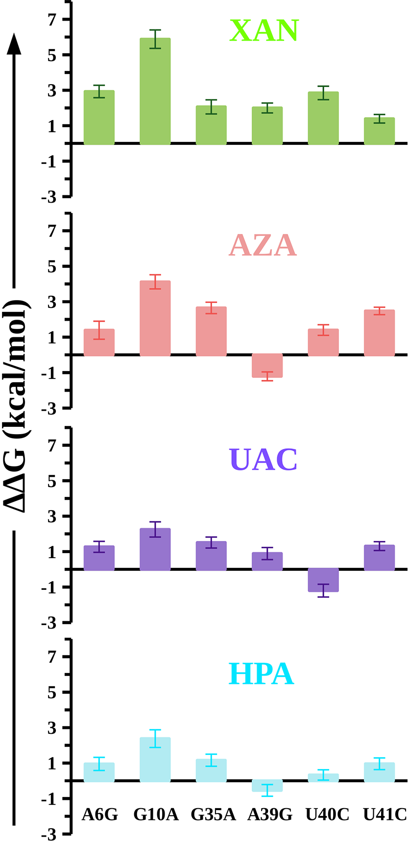
Riboswitches are conserved functional domains in mRNA that almost exclusively exist in bacteria. They regulate the biosynthesis and transport of amino acids and essential metabolites such as coenzymes, nucleobases, and their derivatives by specifically binding small molecules. Due to their ability to precisely discriminate between different cognate molecules as well as their common existence in bacteria, riboswitches have become potential antibacterial drug targets that could deliver urgently needed antibiotics with novel mechanisms of action. In this work, we report the recognition mechanisms of four oxidization products (XAN, AZA, UAC, and HPA) generated during purine degradation by an RNA motif termed the NMT1 riboswitch. Specifically, we investigated the physical interactions between the riboswitch and the oxidized metabolites by computing the changes in the free energy on mutating key nucleobases in the ligand binding pocket of the riboswitch. We discovered that the electrostatic interactions are central to ligand discrimination by this riboswitch. The relative binding free energies of the mutations further indicated that some of the mutations can also strengthen the binding affinities of the ligands (AZA, UAC, and HPA). These mechanistic details are also potentially relevant in the design of novel compounds targeting riboswitches.
Figures


Similar articles
-
Binding site preorganization and ligand discrimination in the purine riboswitch.J Phys Chem B. 2015 Jan 22;119(3):773-82. doi: 10.1021/jp5052358. Epub 2014 Jul 18. J Phys Chem B. 2015. PMID: 25014157
-
Bacterial 2'-Deoxyguanosine Riboswitch Classes as Potential Targets for Antibiotics: A Structure and Dynamics Study.Int J Mol Sci. 2022 Feb 9;23(4):1925. doi: 10.3390/ijms23041925. Int J Mol Sci. 2022. PMID: 35216040 Free PMC article.
-
Insights into xanthine riboswitch structure and metal ion-mediated ligand recognition.Nucleic Acids Res. 2021 Jul 9;49(12):7139-7153. doi: 10.1093/nar/gkab486. Nucleic Acids Res. 2021. PMID: 34125892 Free PMC article.
-
Metabolite recognition principles and molecular mechanisms underlying riboswitch function.Annu Rev Biophys. 2012;41:343-70. doi: 10.1146/annurev-biophys-101211-113224. Annu Rev Biophys. 2012. PMID: 22577823 Free PMC article. Review.
-
[Novel targets for antibiotics discovery: riboswitches].Yao Xue Xue Bao. 2013 Sep;48(9):1361-8. Yao Xue Xue Bao. 2013. PMID: 24358767 Review. Chinese.
Cited by
-
Quantitative Assessment of Energetic Contributions of Residues in a SARS-CoV-2 Viral Enzyme/Nanobody Interface.J Chem Inf Model. 2024 Mar 25;64(6):2068-2076. doi: 10.1021/acs.jcim.3c01933. Epub 2024 Mar 9. J Chem Inf Model. 2024. PMID: 38460144 Free PMC article.
-
Adenine Methylation Enhances the Conformational Flexibility of an RNA Hairpin Tetraloop.J Phys Chem B. 2024 Apr 4;128(13):3157-3166. doi: 10.1021/acs.jpcb.4c00522. Epub 2024 Mar 27. J Phys Chem B. 2024. PMID: 38535997 Free PMC article.
-
Essential Considerations for Free Energy Calculations of RNA-Small Molecule Complexes: Lessons from the Theophylline-Binding RNA Aptamer.J Chem Inf Model. 2025 Jan 13;65(1):223-239. doi: 10.1021/acs.jcim.4c01505. Epub 2024 Dec 19. J Chem Inf Model. 2025. PMID: 39699235
-
Machine-learning prediction of a novel diagnostic model using mitochondria-related genes for patients with bladder cancer.Sci Rep. 2024 Apr 23;14(1):9282. doi: 10.1038/s41598-024-60068-9. Sci Rep. 2024. PMID: 38654047 Free PMC article.
References
-
- Mironov AS; Gusarov I; Rafikov R; Lopez LE; Shatalin K; Kreneva RA; Perumov DA; Nudler E Sensing small molecules by nascent RNA: a mechanism to control transcription in bacteria. Cell 2002, 111, 747–756. - PubMed
-
- Nahvi A; Sudarsan N; Ebert MS; Zou X; Brown KL; Breaker RR Genetic control by a metabolite binding mRNA. Chem. Biol. 2002, 9, 1043–1049. - PubMed
-
- Winkler W; Nahvi A; Breaker RR Thiamine derivatives bind messenger RNAs directly to regulate bacterial gene expression. Nature 2002, 419, 952–956. - PubMed
-
- Blount KF; Breaker RR Riboswitches as antibacterial drug targets. Nat. Biotechnol. 2006, 24, 1558–1564. - PubMed
Publication types
MeSH terms
Substances
Grants and funding
LinkOut - more resources
Full Text Sources
