Review
. 2023 Sep;17(3):768-774.
doi: 10.1007/s12105-023-01575-5. Epub 2023 Jul 24.

Lipoblastoma Arising in the Head and Neck: A Clinicopathologic Analysis of 20 Cases

Affiliations
Review

Lipoblastoma Arising in the Head and Neck: A Clinicopathologic Analysis of 20 Cases

Zahra Aldawood et al. Head Neck Pathol. 2023 Sep.

Abstract

Background: Lipoblastomas (LPBs) are benign adipocytic neoplasms believed to recapitulate the development of embryonal fat.

Methods: We investigated the clinicopathologic and immunohistochemical features of 20 lipoblastomas arising in the head and neck in 18 patients.

Results: Patients included 6 males and 12 females (1:2 ratio) with age at diagnosis ranging from 4 months to 28 years. Tumors occurred more commonly in the neck (12, 66.7%) and less commonly in the forehead, scalp, and tongue (2, 11.1%). Tumor size ranged from 1.4 to 6.0 cm (median 5.0 cm). Two patients, a 4-month-old female and 3-year-old male, had local recurrence of neck tumors at 4 months and 3 years after excision, respectively. Microscopically, tumors had a lobulated growth pattern and consisted of adipocytes at varying stages of differentiation. In addition to the classical histologic features, lipoma-like and myxoid variants constituted 45% of cases. Metaplastic elements, including brown fat and cartilage, were identified in two cases.

Conclusions: LPBs arising in the head and neck region are not uncommon and occurred at a rate of 9% in our cohort. They should be kept in the differential diagnosis when a fatty tumor is encountered in an older child or occurring at an unusual location.

Keywords: Lipoblast; Lipoblastoma; Lipoma; Neck; PLAG1.


Conflict of interest statement

No conflict of interest to disclose.

Figures

Fig. 1
Fig. 1
Common histological features of lipoblastoma. A Grossly, LPB is a circumscribed mass with a yellow vaguely nodular cut surface. B Most tumors exhibited classic features of LPB with an encapsulated lobulated growth pattern and intervening thin fibrous bands. C Focal areas exhibited myxoid stroma. D and E Immature lobules consist of primitive mesenchymal cells admixed with mature adipocytes
Fig. 2
Fig. 2
Lipoma-like lipoblastoma. A Grossly, a lobular brown mass with focal myxoid cut surface (left). B A multilobulated architecture of mature adipocytes with dense intervening fibrous bands. C Immunohistochemical stain for PLAG1 reveals strong nuclear positivity correlating with the presence of gene fusion
Fig. 3
Fig. 3
A 3-year-old female with tongue lipoblastoma. A Grossly, a round mass with a bright yellow surface macroscopically. B Preoperative sagittal MRI image of the head and neck reveals a well-delineated lingual mass (white arrows). C LBP showing lobules of fat in various stages of maturation, surrounded by thick fibrous bands. D Lobules consist of primitive mesenchymal cells admixed with mature adipocytes and a nodule of immature cartilage. EG Multiple foci of cartilage in between the adipocytic lobules consist of primitive mesenchymal cells admixed with mature adipocytes. H Immunohistochemical stain for desmin shows strong positivity within lesional cells. I Immunohistochemical stain for PLAG1 reveals strong nuclear positivity. Note that positive staining includes both primitive cells and metaplastic cartilage
Fig. 4
Fig. 4
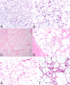
A and B HAS2::PLAG1 fused lipoblastoma in the neck of a 3-year-old female showing marked myxoid stroma with a delicate vascular pattern and numerous lipoblasts. CF Lipoblastoma in the neck of a 3-year-old male showing a classic lobulated pattern with myxoid stroma and variable adipocytic maturation (C, D) in addition to foci of “hibernoma-like” brown adipose tissue with multi-vacuolated adipocytes (E, F)

Similar articles

Cited by

References

    1. Coffin CM, Lowichik A, Putnam A. Lipoblastoma (LPB): a clinicopathologic and immunohistochemical analysis of 59 cases. Am J Surg Pathol. 2009;33(11):1705–1712. doi: 10.1097/PAS.0b013e3181b76462. - DOI - PubMed
    1. Gisselsson D, Hibbard MK, Dal Cin P, Sciot R, Hsi BL, Kozakewich HP, et al. PLAG1 alterations in lipoblastoma: involvement in varied mesenchymal cell types and evidence for alternative oncogenic mechanisms. Am J Pathol. 2001;159(3):955–962. doi: 10.1016/S0002-9440(10)61771-3. - DOI - PMC - PubMed
    1. The WHO Classification of Tumours Editorial Board . WHO classification of tumours soft tissue and bone tumours. 5. Lyon: IARC Press; 2020.
    1. Collins MH, Chatten J. Lipoblastoma/lipoblastomatosis: a clinicopathologic study of 25 tumors. Am J Surg Pathol. 1997;21(10):1131–1137. doi: 10.1097/00000478-199710000-00002. - DOI - PubMed
    1. Fritchie K, Wang L, Yin Z, Nakitandwe J, Hedges D, Horvai A, et al. Lipoblastomas presenting in older children and adults: analysis of 22 cases with identification of novel PLAG1 fusion partners. Mod Pathol. 2020 doi: 10.1038/s41379-020-00696-4. - DOI - PubMed

LinkOut - more resources