Review
. 2023 Jul 24;14(1):136.
doi: 10.1007/s12672-023-00739-7.

WNT ligands in non-small cell lung cancer: from pathogenesis to clinical practice

Affiliations
Review

WNT ligands in non-small cell lung cancer: from pathogenesis to clinical practice

Wanting Xue et al. Discov Oncol. .

Abstract

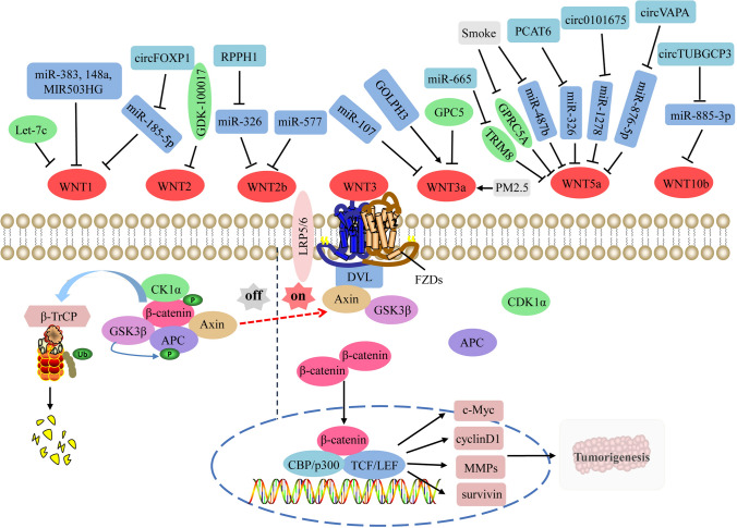
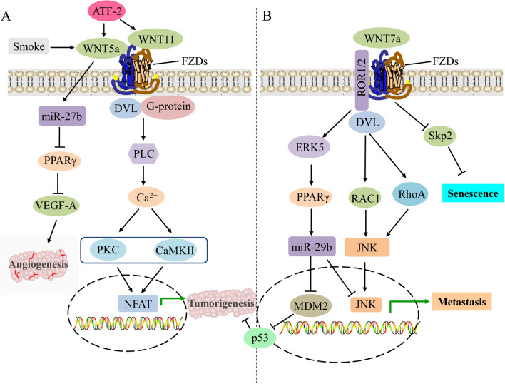
Non-small cell lung cancer (NSCLC) is the malignant tumor with the highest morbidity and leading cause of death worldwide, whereas its pathogenesis has not been fully elucidated. Although mutations in some crucial genes in WNT pathways such as β-catenin and APC are not common in NSCLC, the abnormal signal transduction of WNT pathways is still closely related to the occurrence and progression of NSCLC. WNT ligands (WNTs) are a class of secreted glycoproteins that activate WNT pathways through binding to their receptors and play important regulatory roles in embryonic development, cell differentiation, and tissue regeneration. Therefore, the abnormal expression or dysfunction of WNTs undoubtedly affects WNT pathways and thus participates in the pathogenesis of diseases. There are 19 members of human WNTs, WNT1, WNT2, WNT2b, WNT3, WNT3a, WNT4, WNT5a, WNT5b, WNT6, WNT7a, WNT7b, WNT8a, WNT8b, WNT9a, WNT9b, WNT10a, WNT10b, WNT11 and WNT16. The expression levels of WNTs, binding receptors, and activated WNT pathways are diverse in different tissue types, which endows the complexity of WNT pathways and multifarious biological effects. Although abundant studies have reported the role of WNTs in the pathogenesis of NSCLC, it still needs further study as therapeutic targets for lung cancer. This review will systematically summarize current research on human WNTs in NSCLC, from molecular pathogenesis to potential clinical practice.

Keywords: Non-small cell lung cancer; Therapeutic targets; WNT ligands; WNT signaling pathways.


Conflict of interest statement

The authors have no relevant financial or non-financial interests to disclose.

Figures

Fig. 1
Fig. 1
Regulatory functions of oncogenic WNTs and related upstream regulators on the canonical WNT pathway in NSCLC. In the absence of WNTs, the cytoplasmic β-catenin is phosphorylated by CK1α and GSK3β in destruction complex, followed by ubiquitination by its cognate E3 ligase, SCFβTrCP. When the secreted WNTs are sufficient, they will bind to different FZD receptors (FZDs) and co-receptors LRP5/6, phosphorylate LRP6 and then recruit DVL to the intracellular domain of FZD to form oligomers, then, AXIN and GSK3β dissociate from the destruction complex and bind to DVL. Therefore, β-catenin can be accumulated in the cytoplasm and then translocated into the nucleus to bind to DNA-binding proteins such as TCF/LEF transcription factors, and recruit transcription co-activators CBP and p300 to activate TCF/LEF, thus regulating the transcription of downstream tumor-related target genes that determine cellular proliferation, differentiation and apoptosis, such as c-Myc, cyclinD1, MMPs and survivin. During the onset and progression of NSCLC, Let-7c, miR-383, miR-148a, lncRNA-MIR-503HG, miR-185-5p, miR-326, miR-577, miR-107, miR-326, miR-1278, miR-876-5p, miR-885-3p, cicFOXP1, GPC5, GPRC5A and TRIM8 could block the signaling transduction of the canonical WNT pathway by inhibiting the activities of WNTs or down-regulating their expression, whereas circFOXP1, lncRNA RPPH1, GOLPH3, smoke, PM2.5, lncRNA-PCAT6, circ101675, circVAPA and circTUBGCP3 could activate the canonical WNT pathway by over-activating WNTs or up-regulating their expression. APC adenomatosis polyposis coli, β-TRCP β-transducin repeat-containing protein, CBP CREB-binding protein, CK1α casein kinase 1α, DVL disheveled, FOXP1, forkhead box protein P1, FZDs frizzleds, GDK-100017 2,3,6-trisubstituted quinoxaline derivative, GOLPH3 Golgi phosphoprotein 3, GPC5 glypican-5, GPRC5A G protein coupled receptor family C group 5 type A, GSK3β glycogen synthase kinase 3β, LEF lymphoid enhancer-binding factor, MMPs matrix metalloproteinases, NSCLC non-small cell lung cancer, PCAT6 prostate cancer-associated transcript 6, RPPH1 ribonuclease P RNA component H1, TCF T-cell factor, TRIM8 tripartite motif-containing 8, TUBGCP3 gamma tubulin complex component 3
Fig. 2
Fig. 2
Regulatory functions of anti-cancer WNTs by activating the noncanonical WNT pathways in NSCLC. A WNT5a and WNT11 bind to FZDs and G protein subunit to recruit DVL, which triggers PLC activity and promotes intracellular Ca2+ release, and further activates calcium-dependent PKC and CAMKII signaling pathways, thus to regulate the expression of downstream genes to promote the proliferation and invasion of NSCLC cells by inducing the accumulation of nuclear NFAT transcription factors. WNT5a also inhibits VEGF-A-induced endothelial cell migration and motility to promote angiogenesis by inducing miR-27b and direct consequence of PPARγ reduction in NSCLC cells. Smoke and ATF-2 could activate the noncanonical WNT pathway by up-regulating the expression of WNT5a and WNT11. B The β-catenin-independent Wnt/PCP/JNK pathway is initiated by the cumulative binding of WNT7a to the ROR1/2-FZDs complex, then DVL is activated to bind to some small Rho GTPases such as RAC1 and RhoA to trigger JNK. This results in the inhibition of transformed cell growth but enhancement of migration and invasion of NSCLC cells. The binding of WNT7a to FZD9 inhibits cellular transformation and proliferation of NSCLC cells by activating the tumor suppressor PPARγ via the ERK5-dependent pathway, thus to relieve the inhibitory effect of MDM2 on p53 tumor suppressor pathway by inducing the expression of anti-cancer miR-29b, and promotes epithelial differentiation through activating JNK pathway and the resultant upregulation of cadherins. Wnt7a also triggers the cellular senescence of lung cancer via the inactivation of Skp2, a key negative regulator of cellular senescence. CAMKII calmodulin-dependent protein kinase II, DVL disheveled, ERK5 extracellular signal-regulated kinase 5, JNK JUN N-terminal kinase, MDM2 murine double minute 2, NFAT nuclear factor of activated T cells, NSCLC non-small cell lung cancer, PCP planar cell polarity, PKC protein kinase C, PLC phospholipase C, PPARγ peroxisome proliferator–activated receptor-γ, RAC1 Rac family small GTPase 1, ROR1/2 receptor tyrosine kinase-like orphan receptor 1/2, Skp2 S-phase kinase-associated protein-2, VEGF-A vascular endothelial growth factor-A

Similar articles

Cited by

References

    1. Sung H, Ferlay J, Siegel RL, Laversanne M, Soerjomataram I, Jemal A, et al. Global Cancer Statistics 2020: GLOBOCAN estimates of incidence and Mortality Worldwide for 36 cancers in 185 countries. CA Cancer J Clin. 2021;71(3):209–49. doi: 10.3322/caac.21660. - DOI - PubMed
    1. Minna JD, Roth JA, Gazdar AF. Focus on lung cancer. Cancer Cell. 2002;1(1):49–52. doi: 10.1016/S1535-6108(02)00027-2. - DOI - PubMed
    1. Stewart DJ. Tumor and host factors that may limit efficacy of chemotherapy in non-small cell and small cell lung cancer. Crit Rev Oncol Hematol. 2010;75(3):173–234. doi: 10.1016/j.critrevonc.2009.11.006. - DOI - PMC - PubMed
    1. Garon EB, Hellmann MD, Rizvi NA, Carcereny E, Leighl NB, Ahn MJ, et al. Five-year overall survival for patients with Advanced non–small-cell lung Cancer treated with pembrolizumab: results from the phase I KEYNOTE-001 study. J Clin Oncol. 2019;37(28):2518–27. doi: 10.1200/JCO.19.00934. - DOI - PMC - PubMed
    1. Wood SL, Pernemalm M, Crosbie PA, Whetton AD. The role of the tumor-microenvironment in lung cancer-metastasis and its relationship to potential therapeutic targets. Cancer Treat Rev. 2014;40(4):558–66. doi: 10.1016/j.ctrv.2013.10.001. - DOI - PubMed

LinkOut - more resources