. 2023 Oct;12(5):2641-2655.
doi: 10.1007/s40123-023-00769-y. Epub 2023 Jul 24.

Impact of Latanoprost Antiglaucoma Eyedrops and Their Excipients on Toxicity and Healing Characteristics in the Ex Vivo Eye Irritation Test System

Affiliations

Impact of Latanoprost Antiglaucoma Eyedrops and Their Excipients on Toxicity and Healing Characteristics in the Ex Vivo Eye Irritation Test System

Claudia Panfil et al. Ophthalmol Ther. 2023 Oct.

Abstract

Introduction: Corneal epithelial toxicity and delayed healing process have already been attributed to preservatives or some excipients. We study the effects of galenic components in antiglaucoma drugs such as benzalkonium chloride (BAC) or surfactants like macrogolglycerol hydroxystearate 40 (MGHS 40) on corneal toxicity in an ex vivo system mimicking chronic use.

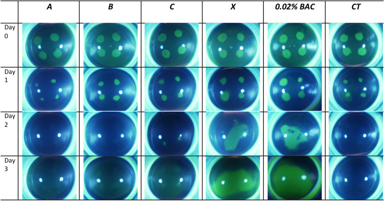
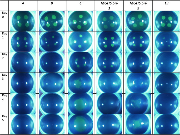
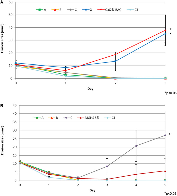
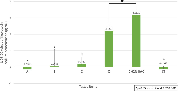
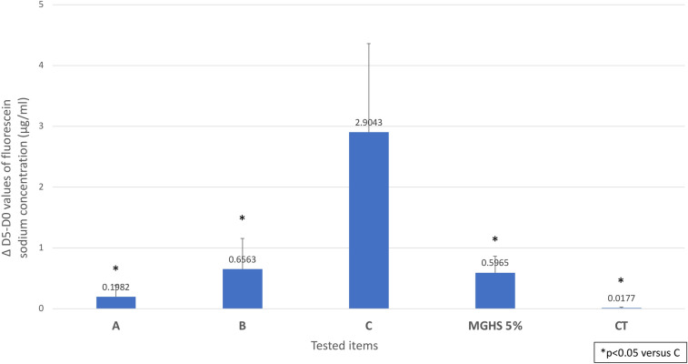
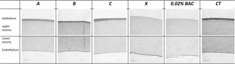
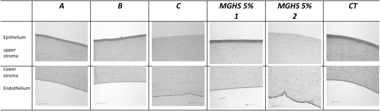
Methods: Latanoprost-containing eyedrops are available with and without preservatives on the market. Unpreserved, they are available in different formulations with various excipients like MGHS at different concentrations (0%, 2.5%, and 5%). We studied these in the ex vivo bioreactor (EVEIT) on initially injured rabbit corneas. The drugs were applied six times daily for observation periods of 3 or 5 days. BAC, 5% MGHS 40 solution, and 0.18% hyaluronic acid served as controls. Macroscopic photographic, biochemical methods and corneal integrity quantification were used for evaluation. Toxicity was assessed by measuring wound healing and corneal fluorescein sodium permeability and was confirmed by histology studies.

Results: The BAC-preserved formulation resulted in high corneal toxicity, which was expected. Interestingly, the preservative-free (PF) formulation containing 5% MGHS 40, carbomer, macrogol 4000, and sorbitol showed the highest corneal toxicity, followed by the control formulation with equal MGHS 40 concentration, which presented significantly less damage. No toxicity was shown by eyedrops containing 2.5% MGHS 40 or salts only.

Conclusion: Our study demonstrates a significant corneal toxicity of certain formulations of PF antiglaucoma ophthalmic drugs containing 5% MGHS 40 with other excipients compared to other formulations with lower MGHS 40 concentrations (2.5% or 0%), or even compared to the solution containing 5% MGHS alone. This suggests a concentration-dependent toxicity of MGHS 40, especially in interaction with other excipients, which may increase its epithelial toxicity, and that has to be considered in clinical glaucoma therapy. Further single-component formulation trials are needed to support this interpretation.

Keywords: BAC; Corneal healing; Cytotoxicity; Excipient; Ex vivo model; Glaucoma medication; Latanoprost; Macrogolglycerol hydroxystearate 40; Preservative.


Conflict of interest statement

Claudia Panfil and Norbert Schrage have nothing to disclose. Laure Chauchat, Camille Guerin, Hayetta Rebika and Marwan Sahyoun declare working for Horus Pharma at the time of completion of the manuscript.

Figures

Fig. 1
Fig. 1
Microscopic evaluation of the epithelial healing process of the fluorescein stained corneas during 3 days observation period (n = 3 corneas per test item). Healing was observed from day 1 for the control reference CT, completed by day 2, closely followed by latanoprost formulations A and B with stable epithelial closure at day 3. After initial healing, the control 0.02% BAC and BAC-preserved latanoprost formulation X showed strong epithelial reopening from day 2 to day 3. Group C showed delayed healing at day 2, not completed at day 3. BAC benzalkonium chloride, MGHS macrogolglycerol hydroxystearate 40
Fig. 2
Fig. 2
Representative fluorescein-stained microscopical images of the corneal healing process during experimental period of 5 days with drug application six times daily (n = 5 corneas per test item). All five corneas per test item are shown in detail in Figs. S1–S5 in the supplementary material. Healing was observed from day 1 for the control reference CT, completed by day 2, closely followed by latanoprost formulations A and B with stable epithelial closure at day 3. Group C showed delayed healing at day 2, not completed at day 3 and significant reopening of the lesions up to day 5. For the corneas treated with MGHS 5% alone, different healing processes were observed throughout the study period, ranging from complete and stable healing (MGHS 5% 1) to delayed healing with residual lesions and an increase in lesion size compared to baseline (MGHS 5% 2). MGHS macrogolglycerol hydroxystearate 40
Fig. 3
Fig. 3
Graphical illustration of the corneal healing process under six times daily drug application over 3 days (a) with n = 3 corneas or 5 days (b) with n = 5 corneas per test item. The mean corneal erosion sizes in mm2 are plotted against time (experimental days). We observe a final significant (p < 0.05) reopening of the epithelium compared to baseline for formulations X and C and the control 0.02% BAC. BAC benzalkonium chloride, MGHS macrogolglycerol hydroxystearate 40
Fig. 4
Fig. 4
Corneal integrity changes of all corneas (n = 3 each test item) after 3 experimental days of fluorescein sodium permeation through the cornea. Fluorescein permeability Δ value was significantly higher for BAC-preserved latanoprost formulation X and the control 0.02% BAC (p < 0.05) than for A, B, C, and CT. No significant difference was seen between formulation X and BAC. BAC benzalkonium chloride
Fig. 5
Fig. 5
Corneal integrity changes of all corneas (n = 5 each test item) after 5 experimental days of fluorescein sodium permeation through the cornea. The ΔD5–D0 values were significantly higher for C compared to A, B, and MHGS 5% (p < 0.05) highlighting a reduction in corneal integrity due to test substance application six times daily. No statistical difference was observed between the ΔD5–D0 values of A, B, and CT. MGHS macrogolglycerol hydroxystearate 40
Fig. 6
Fig. 6
Graphical analysis of the daily measured lactate and glucose values as an indicator of metabolic activity during test substance application over 3 days (a, b) or 5 days (c, d). Standard deviations are integrated. There is an increase in lactate metabolism measured for C, X, and 0.02% BAC control, but only for formulation X were latanoprost values significantly different (p < 0.05) at day 3 compared to baseline (a). For the 5-day testing, lactate levels remained in physiological range, with a significant increase for C and MGHS 5% (p < 0.05) compared to CT (c). In both studies, glucose levels remained in physiological range (b, d). BAC benzalkonium chloride, MGHS macrogolglycerol hydroxystearate 40
Fig. 7
Fig. 7
Histologic evaluation after 3 days drip application of the eye drugs A, B, C, and X and controls. Scale bar 100 µm. Formulations A and B and CT show complete healing of the epithelial layer, dense stroma, and regularly arranged keratocytes. Moreover, Descemet’s membrane and the endothelial layer were intact without any structural damage. For all corneas treated with substance C, re-epithelization with slight corneal edema was observed. Histology of the BAC-preserved latanoprost formulation X showed severe superficial corneal damage with complete epithelial cell loss, distinct local edema, and keratocyte loss with still intact Descemet and endothelial layers. The same result was seen on day 3 in the 0.02% BAC control group, even with detached Descemet’s membrane in one of three corneas. Hyaluronic acid-containing control CT shows perfectly and stably healed multilayered epithelium, intact stroma, Descemet, and endothelium. BAC benzalkonium chloride
Fig. 8
Fig. 8
Histologic evaluation after prolonged 5-day treatment with ophthalmic drugs A, B, and C and the control MGHS 5% (macrogolglycerol hydroxystearate 40) and hyaluronic acid CT. Scale bar 100 µm. Histological findings confirmed microscopic data for all test substances. Formulations A and B and CT show complete healing of the epithelial layer, dense stroma, and regularly arranged keratocytes. Moreover, Descemet’s membrane and the endothelial layer were intact without any structural damage. For corneas treated with substance C, the five corneas showed a total loss of epithelial cells, distinct stromal edema, severe loss of keratocytes, and a damaged endothelium detached from Descemet’s membrane. This formulation demonstrates very clearly the toxicological effects of eyedrops as a function of the observation period. Findings were similar for two out of five corneas treated with MGHS 5% (Fig. MGHS 5% 2) in contrast to the other three corneas of this group whose epithelium was histologically thin in places but closed (Fig. MGHS 5% 1). Hyaluronic acid-containing control CT in both studies shows perfectly and stably healed multilayered epithelium, intact stroma, Descemet, and endothelium

Similar articles

Cited by

References

    1. European Glaucoma Society. European Glaucoma Society terminology and guidelines for glaucoma, 5th edition. Br J Ophthalmol. 2021;105:1–169. - PubMed
    1. Boimer C, Birt CM. Preservative exposure and surgical outcomes in glaucoma patients: the PESO study. J Glaucoma. 2013;22:730–735. doi: 10.1097/IJG.0b013e31825af67d. - DOI - PubMed
    1. Konstas AG, Labbé A, Katsanos A, et al. The treatment of glaucoma using topical preservative-free agents: an evaluation of safety and tolerability. Expert Opin Drug Saf. 2021;20:453–466. doi: 10.1080/14740338.2021.1873947. - DOI - PubMed
    1. Hakkarainen JJ, Reinisalo M, Ragauskas S, Seppänen A, Kaja S, Kalesnykas G. Acute cytotoxic effects of marketed ophthalmic formulations on human corneal epithelial cells. Int J Pharm. 2016;511:73–78. doi: 10.1016/j.ijpharm.2016.06.135. - DOI - PubMed
    1. Smedowski A, Paterno JJ, Toropainen E, Sinha D, Wylegala E, Kaarniranta K. Excipients of preservative-free latanoprost induced inflammatory response and cytotoxicity in immortalized human HCE-2 corneal epithelial cells. J Biochem Pharmacol Res. 2014;2:175–184. - PMC - PubMed

LinkOut - more resources