. 2023 Dec 1;74(22):7034-7044.
doi: 10.1093/jxb/erad284.

A roadmap of haustorium morphogenesis in parasitic plants

Affiliations

A roadmap of haustorium morphogenesis in parasitic plants

Gwendolyn K Kirschner et al. J Exp Bot. .

Abstract

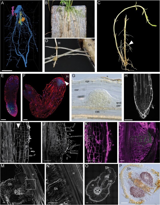
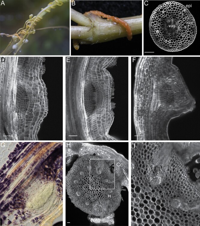
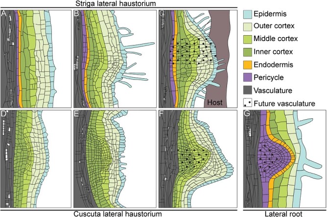
Parasitic plants invade their host through their invasive organ, the haustorium. This organ connects to the vasculature of the host roots and hijacks water and nutrients. Although parasitism has evolved independently in plants, haustoria formation follows a similar mechanism throughout different plant species, highlighting the developmental plasticity of plant tissues. Here, we compare three types of haustoria formed by the root and shoot in the plant parasites Striga and Cuscuta. We discuss mechanisms underlying the interactions with their hosts and how different approaches have contributed to major understanding of haustoria formation and host invasion. We also illustrate the role of auxin and cytokinin in controlling this process.

Keywords: Cuscuta; Striga; Auxin; cytokinin; haustoria; host; invasion; lateral roots; parasitic plant.


Conflict of interest statement

The authors declare no conflict of interest.

Figures

Fig. 1.
Fig. 1.
Terminal and lateral haustorium development in Striga hermonthica. (A) Micro-CT 3D reconstruction and post-processed image segmentation depicting the association of Striga seedlings (orange, yellow, and purple) attached to the rice roots (blue). (B) Macrophotograph showing Striga plants grown on rice as a host plant on top of nylon mesh. (C) Macrophotograph showing the Striga plant isolated from the host plants. Note the emerging adventitious roots (arrowhead). (D) Attachment of Striga adventitious roots to the host roots by lateral haustoria. (E and F) Confocal image showing the root meristem of Striga seedlings directly after germination (E) and before attachment to the host root (F). Roots were stained with modified pseudo-Schiff propidium iodide (mPS-PI) (red) (Truernit et al., 2008), dividing cells were visualized by 5-ethynyl-2ʹ-deoxyuridine (EdU) staining (green), and nuclei were stained with Hoechst (blue); the arrowhead points to haustorial hairs. (G) Lateral root primordium of Striga roots stained with Lugol, cleared with chloral hydrate, and visualized with Nomarski microscopy. (H) Confocal image of a Striga lateral root tip; cell walls stained with mPS-PI. (I and J) Confocal images of longitudinal vibratome sections of a Striga root developing a lateral haustorium; cell walls and starch granules were stained with mPS-PI; the arrowhead points to periclinal cell divisions in the inner cortex upon haustorium initiation. (K and L) Confocal longitudinal sections showing cell divisions in developing Striga lateral haustoria. EdU-stained nuclei are green and cell walls stained with SCRI Renaissance are in magenta. (M) Longitudinal section of a Striga lateral haustorium during attachment to the host plant; (N) magnification of the area marked in (M). (O) Cross-sections of a Striga lateral haustorium attached to a host root; cell walls stained with mPS-PI. (P) Starch granules accumulation in the Striga lateral haustorium, visualized by Lugol’s staining. cor, cortex; end, endodermis; epi, epidermis; H, host; HB, hyaline body; per, pericycle; Sh, Striga hermonthica; vas, vasculature; XB, xylem bridge. Scale bars 20 μm (A, B), 50 μm (E, F, G, H, I, J, K, L, M, N, O, P).
Fig. 2.
Fig. 2.
Cuscuta haustorium development. (A and B) Cuscuta plants grown around Sphagneticola trilobata as host plants. (C) Confocal image of a vibratome cross-section of a Cuscuta stem; cell walls are stained with SCRI Renaissance. (D–F) Longitudinal vibratome sections of a Cuscuta stem developing a haustorium; cell walls stained are with SCRI Renaissance. (G) Accumulation of starch granules during haustorium formation, visualized by Lugol’s staining. (H and I) Vibratome cross-sections of the host stem with Cuscuta attached to it with haustoria; cell walls stained with mPS-PI (Truernit et al., 2008); (I) magnification of the area marked in (H). Cc, Cuscuta campestris; H, host; cor, cortex, epi, epidermis, vas, vasculature; scale bars 50 μm.
Fig. 3.
Fig. 3.
Organogenesis of lateral haustoria and lateral roots. (A–C) Striga lateral haustorium development. (D–F) Cuscuta lateral haustorium development. (G) Lateral root initiation in Arabidopsis; tissues are color-coded according to the key.

References

    1. Aoki N, Cui S, Yoshida S.. 2022. Cytokinins induce prehaustoria coordinately with quinone signals in the parasitic plant Striga hermonthica. Plant and Cell Physiology 63, 1446–1456. - PubMed
    1. Benková E, Michniewicz M, Sauer M, Teichmann T, Seifertová D, Jürgens G, Friml J.. 2003. Local, efflux-dependent auxin gradients as a common module for plant organ formation. Cell 115, 591–602. - PubMed
    1. Bernal-Galeano V, Beard K, Westwood JH.. 2022. An artificial host system enables the obligate parasite Cuscuta campestris to grow and reproduce in vitro. Plant Physiology 189, 687–702. - PMC - PubMed
    1. Berner DK, Kling JG, Singh BB.. 1995. Striga research and control—a perspective from Africa. Plant Disease 79, 652–660.
    1. Cai T, Babiker AG, Ejeta G, Butler LG.. 1993. Morphological response of witchweed (Striga asiatica) to in vitro culture. Journal of Experimental Botany 44, 1377–1384.

Publication types