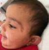
Case Report: Extensive Facial Cutaneous Leishmaniasis in a Neonate
- PMID: 37487561
- PMCID: PMC10484260
- DOI: 10.4269/ajtmh.23-0177
Case Report: Extensive Facial Cutaneous Leishmaniasis in a Neonate
Abstract
Cutaneous leishmaniasis (CL) is a skin infection caused by various species of the Leishmania parasite and is spread by the bite of an infected female sandfly. In southern Israel, CL caused by Leishmania major is endemic. Cutaneous leishmaniasis is considered a self-limiting disease, characterized by progressive, long-lasting nodulo-ulcerative skin lesions, which usually resolve in several months to years, and leads to scarring, cosmetic disfigurement, and future stigmatization. Although CL is a common disease among children, reports of CL in children younger than 1 year are rare. We present a case of extensive facial CL in an infant whose initial lesions appeared only 25 days after birth. The patient was treated with intravenous liposomal amphotericin B. Two months later, marked improvement was seen, with complete resolution of the inflammation and atrophic scar formation. To our knowledge, this is the earliest age of CL published to date.
Figures


References
-
- Horev A, Sagi O, Zur E, Ben-Shimol S, 2023. Topical liposomal amphotericin B gel treatment for cutaneous leishmaniasis caused by Leishmania major: a double-blind, randomized, placebo-controlled, pilot study. Int J Dermatol 62: 40–47. - PubMed
-
- Noyman Y, Levi A, Ben Amitai D, Reiss-Huss S, Sabbah F, Hodak E, Mimouni T, Friedland R, 2022. Treating pediatric cutaneous Leishmania tropica with systemic liposomal amphotericin B: a retrospective, single-center study. Dermatol Ther (Heidelb) 35: e15185. - PubMed
-
- Erat T, An I, 2022. Treatment of pediatric cutaneous leishmaniasis with liposomal amphotericin B. Dermatol Ther (Heidelb) 35: e15706. - PubMed
Publication types
MeSH terms
Substances
LinkOut - more resources
Full Text Sources

