. 2023 Jul;11(7):e006921.
doi: 10.1136/jitc-2023-006921.

Autologous humanized PDX modeling for immuno-oncology recapitulates features of the human tumor microenvironment

Affiliations

Autologous humanized PDX modeling for immuno-oncology recapitulates features of the human tumor microenvironment

Michael Chiorazzi et al. J Immunother Cancer. 2023 Jul.

Abstract

Background: Interactions between immune and tumor cells are critical to determining cancer progression and response. In addition, preclinical prediction of immune-related drug efficacy is limited by interspecies differences between human and mouse, as well as inter-person germline and somatic variation. To address these gaps, we developed an autologous system that models the tumor microenvironment (TME) from individual patients with solid tumors.

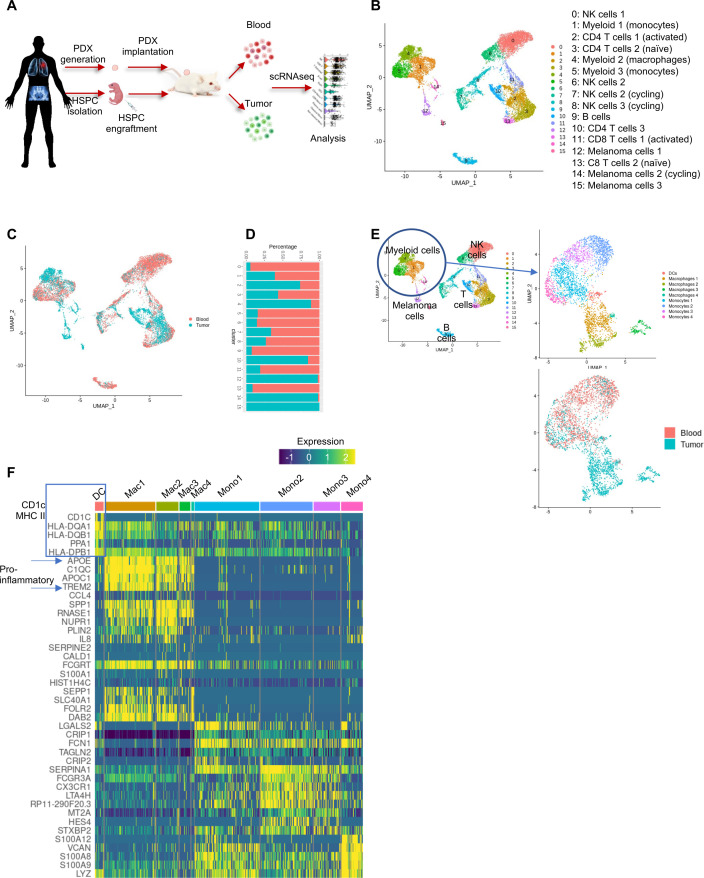
Method: With patient-derived bone marrow hematopoietic stem and progenitor cells (HSPCs), we engrafted a patient's hematopoietic system in MISTRG6 mice, followed by transfer of patient-derived xenograft (PDX) tissue, providing a fully genetically matched model to recapitulate the individual's TME. We used this system to prospectively study tumor-immune interactions in patients with solid tumor.

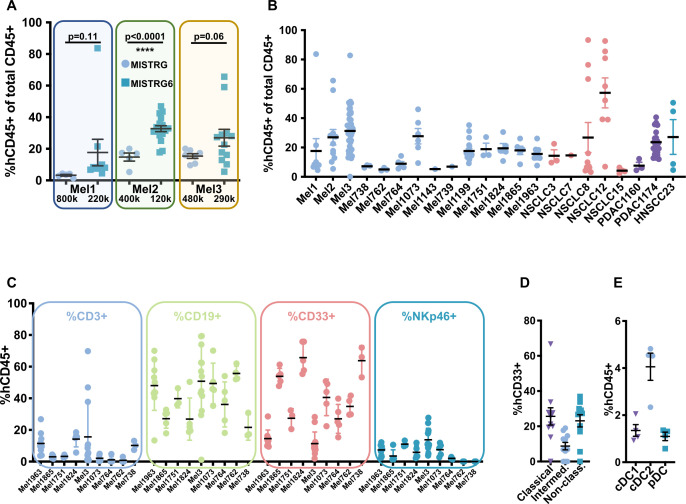
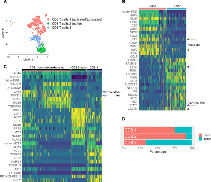
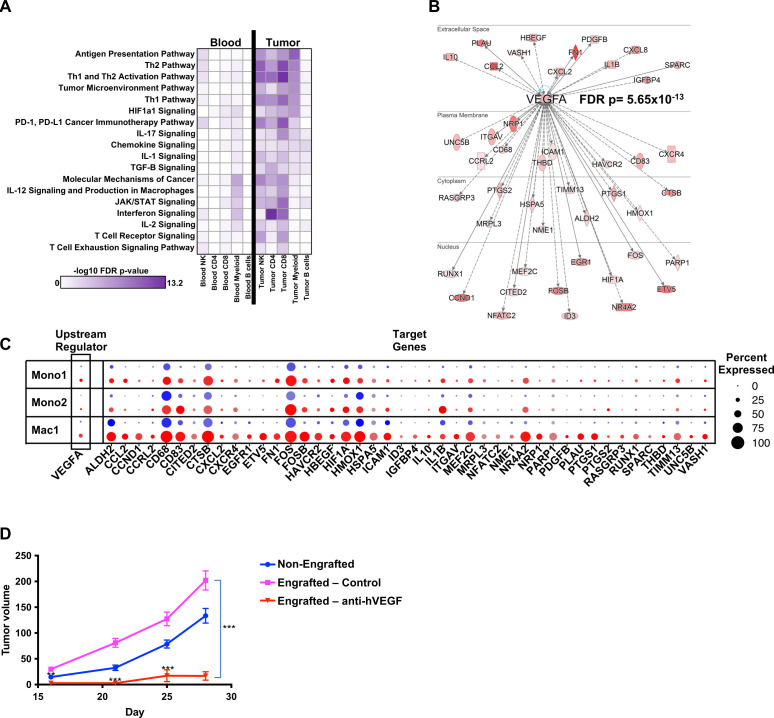
Results: Autologous PDX mice generated innate and adaptive immune populations; these cells populated the TME; and tumors from autologously engrafted mice grew larger than tumors from non-engrafted littermate controls. Single-cell transcriptomics revealed a prominent vascular endothelial growth factor A (VEGFA) signature in TME myeloid cells, and inhibition of human VEGF-A abrogated enhanced growth.

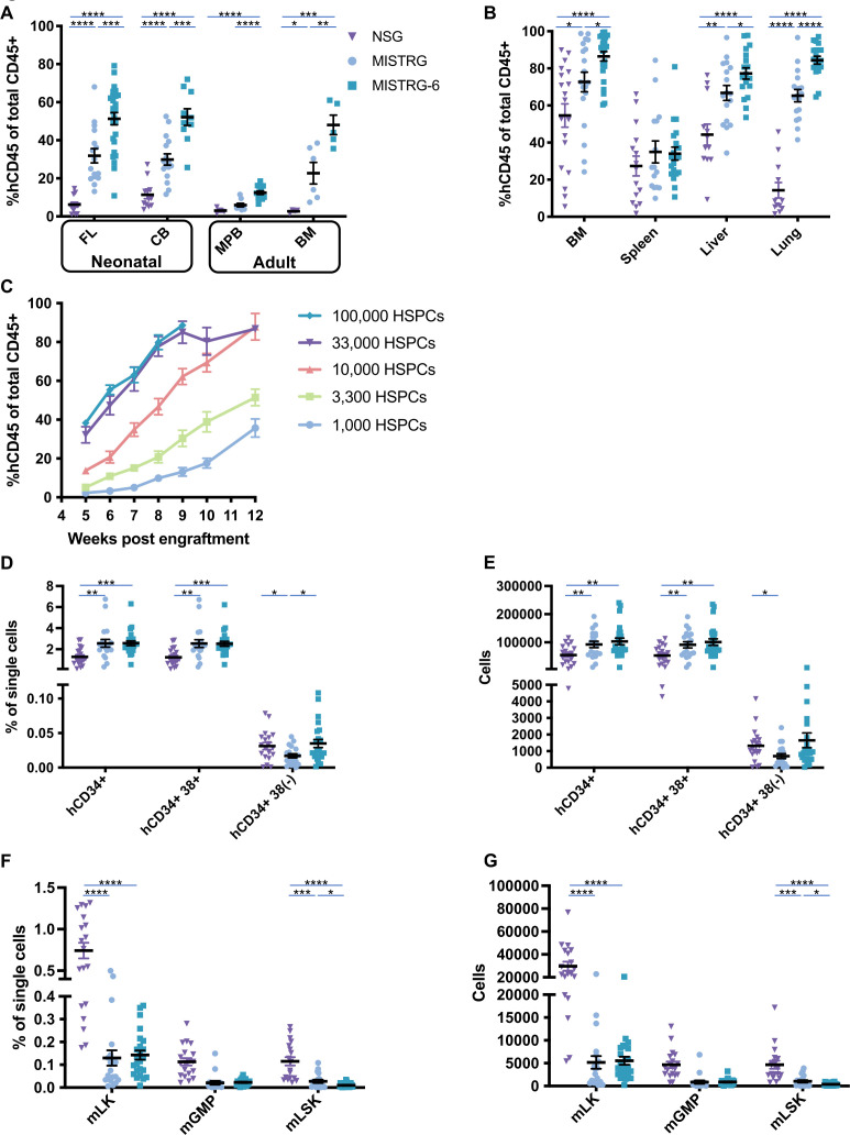
Conclusions: Humanization of the interleukin 6 locus in MISTRG6 mice enhances HSPC engraftment, making it feasible to model tumor-immune interactions in an autologous manner from a bedside bone marrow aspirate. The TME from these autologous tumors display hallmarks of the human TME including innate and adaptive immune activation and provide a platform for preclinical drug testing.

Keywords: Immunity, Innate; Immunotherapy; Inflammation; Macrophages; Tumor Microenvironment.


Conflict of interest statement

Competing interests: HHMI lab heads have previously granted a non-exclusive CC BY 4.0 license to the public and a sublicensable license to HHMI in their research articles. Pursuant to those licenses, the author-accepted manuscript of this article can be made freely available under a CC BY 4.0 license immediately upon publication. RF is an advisor to GlaxoSmithKline, EvolveImmune, and Ventus Therapeutics.

Figures

Figure 1
Figure 1
Humanization of the IL-6 locus enhances human hematopoietic engraftment in MISTRG6 mice. (A) Human hematopoietic engraftment (percent of hCD45+ cells as a proportion of total hCD45+ and mCD45+ cells) in peripheral blood of NSG (triangles), MISTRG (circles) and MISTRG6 (squares) mice engrafted with equal numbers of CD34+ HSPCs from indicated sources; *p<0.05, **p<0.01, ***p<0.001, ****p<0.0001, unpaired parametric t-test; bars indicate mean and SEM); each dot represents a single mouse. 1–3 day-old pups were irradiated with 150 rads, intrahepatically injected with 100,000 FL, 50,000 CB, or 100,000–180,000 adult PB CD34+ cells. Blood engraftment was measured at 5–7 weeks post engraftment. (B) Human hematopoietic engraftment (%hCD45+ of total CD45+) in indicated tissues of NSG, MISTRG and MISTRG6 mice engrafted with equal numbers of CD34+ HSPCs. (C) Longitudinal analysis of human CD45+ cells in peripheral blood of MISTRG6 mice engrafted with varying HSPC numbers as indicated; n=6–11 for each group. (D) Percentage among single cells of hCD34+, hCD34+hCD38+, and hCD34+hCD38(−) cells detected in BM of NSG, MISTRG and MISTRG6 mice engrafted with equal numbers of hCD34+ HSPCs. (E) Absolute numbers of cells in BM of mice in (D). (F) Percentage among single cells of mouse mLK, mGMP and mLSK cells detected by flow cytometry in BM of NSG, MISTRG and MISTRG6 mice engrafted with equal numbers of CD34+ HSPCs. (G.) Absolute numbers of cells from BM of mice in (F). BM, bone marrow; CB, cord blood; FL, fetal liver; HSPCs, hematopoietic stem and progenitor cells; IL, interleukin; MPB, mobilized peripheral blood; PB, peripheral blood.
Figure 2
Figure 2
Improved engraftment in MISTRG6 compared with MISTRG mice of HSPCs from patients with solid tumor, with human innate and adaptive immune cells represented. (A) Analysis at 8 weeks of age of peripheral blood of MISTRG (circles) and MISTRG6 (squares) mice engrafted with HSPCs from the indicated patients with melanoma with the HSPC dose shown below; note improved human engraftment levels despite fewer HSPCs introduced in MISTRG6 compared with MISTRG. (B) Human hematopoietic engraftment of MISTRG6 mice with 100–250 k HSPCs derived from adult patients with melanoma (Mel), non-small cell lung cancer (NSCLC), pancreatic adenocarcinoma (PDAC), and squamous cell carcinoma of the head and neck (HNSCC); each dot represents a single mouse. (C) Percentage of human T cells (CD3+), B cells (CD19+), myeloid cells (CD33+) and NK cells (NKp46+) out of total human hematopoietic cells in peripheral blood of autologously engrafted mice from the indicated patients. (D) Proportions of human monocyte subsets (CD14+CD16(−) classical, triangles; CD14+CD16+ intermediate, circles; CD14(−)CD16+non-classical, squares) of total CD33+ myeloid cells in peripheral blood of Mel1963 autologously engrafted MISTRG6 mice. (E) Proportions of human dendritic cell subsets (HLA-DR+CD11clowCD141+ cDC1, triangles; CD11c+CD1c+ cDC2, circles; CD14(−)CD11c(−)CD303+ pDC, squares) of total hCD45+ cells in spleens of Mel1963 autologously engrafted MISTRG6 mice. HSPCs, hematopoietic stem and progenitor cells; NK, natural killer.
Figure 3
Figure 3
Autologous engraftment enhances PDX growth and human immune cell infiltrate demonstrates tumor-immune interactions. (A) PDX growth in MISTRG6 littermates engrafted (magenta) or non-engrafted (blue) with autologous HSPCs from indicated patients (see online supplemental figure 2 for comprehensive data). (B) H&E (left) and hCD45 immunohistochemical (right) staining of tumors harvested from Mel1073 PDX grown in non-engrafted hosts (top), autologously engrafted MISTRG6 hosts and the patient’s primary tumor (bottom). (C) Sections of Mel1199 PDX tumor grown in autologously engrafted host stained for human infiltrating immune cells; top panel blue=DAPI, cyan=hCD3, green=hCD14, red=hHLA DR, white=melanoma antigen, scale bar=100 um. Bottom panel shows the same markers without melanoma antigen staining. Green arrows highlight HLA-DR+CD14+ macrophages while red arrows highlight HLA-DR+CD14(−) antigen-presenting cells. Cyan arrow highlights T cell-macrophage interaction. Scale bar=50 um. (D.) Number of somatic mutations (compared with germline reference) shared between the indicated samples: Mel738 patient’s surgical resection sample = 1o Met; PDX tumors from non-engrafted mice (lacking human immune cells) = NE tumor A and B; PDX tumors from mice with autologous engraftment=HuMo A and B; cell line derived from the patient’s tumor=cell line. Note that most mutations (225) are shared among all samples. HNSCC, squamous cell carcinoma of the head and neck; HSPCs, hematopoietic stem and progenitor cells; Mel, melanoma; NSCLC, non-small cell lung cancer; PDX, patient-derived xenograft.
Figure 4
Figure 4
Single cell genomics reveals multiple human immune cell types in tumors and blood of autologous MISTRG6 PDX mice, including innate immune cell types present in the TME. (A) Schematic representation of scRNAseq experiments. (B) UMAP embedding displaying unsupervised clustering of 14,603 human cells from blood and melanoma PDX of autologous MISTRG6 mice. Cell types were identified by marker genes and identities are listed. (C) UMAP embedding displaying tissue of origin for cells in A; red cells are derived from blood libraries, blue cells from tumor libraries. (D) Proportions of cluster representation in blood versus tumor scRNAseq libraries. (E) Re-clustering of myeloid cells reveals substructure of nine clusters including DCs, macrophages and monocytes in differential tissue representation as indicated in the bottom panel. (F) Heatmap indicating expression of top differentially expressed genes between each cluster, highlighting presence of human DCs in TME and pro-inflammatory macrophage subtypes. DCs, dendritic cells; HSPCs, hematopoietic stem and progenitor cells; NK, natural killer; PDX, patient-derived xenograft; scRNAseq, single-cell RNA sequencing; TME, tumor microenvironment; UMAP, Uniform Manifold Approximation and Projection.
Figure 5
Figure 5
CD8 T cells circulating in the blood of autologous MISTRG6 mice display features of naïve states while those in the TME express markers of activation and exhaustion. (A) Re-clustering of CD8 T cells reveals substructure of three clusters. (B) Differentially-expressed genes between CD8 T cells in blood versus melanoma PDX display features of naïve (blood) and activated (tumor) states. (C) Heatmap indicating expression of top differentially expressed genes between each cluster, highlighting activated/exhausted-like phenotype of CD8 1 cluster. (D) Cluster representation of CD8 T cell subclusters in tissues, demonstrating over-representation of activated/exhausted-like CD8 T cells in the tumor microenvironment of autologous mice. PDX, patient-derived xenograft; TME, tumor microenvironment; UMAP, Uniform Manifold Approximation and Projection.
Figure 6
Figure 6
Immune cells in the TME display gene signatures associated with immune activation and signaling, including VEGF-A signaling, and blockade of this molecule abrogates enhanced tumor growth in engrafted autologous PDX mice. (A) Canonical pathway representation in indicated cell types from blood (left) and melanoma tumor PDX (right). (B) Upstream pathway analysis identifies VEGFA and its target genes as highly represented in the TME; red shading indicates VEGFA target gene expression level in the TME. (C) VEGFA and its target genes are over-represented in the TME (red) of myeloid gene clusters (monocyte 1, monocyte 2, macrophage 1) compared with blood (blue); size of split dot plot circle indicates percent expression of each gene among cells of that cluster, while intensity of color indicates level of expression. (D) Treatment of autologously engrafted MISTRG6 Mel2 PDX mice with bevacizumab, a clinical anti-hVEGF-A therapy, significantly reduces tumor growth compared with autologous HSPC-engrafted control (asterisks indicate comparison between drug-treated and control engrafted mice); n=13 non-engrafted, n=4 engrafted control, n=5 bevacizumab-treated engrafted mice. FDR, false discovery rate; HSPC, hematopoietic stem and progenitor cell; PDX, patient-derived xenograft; TME, tumor microenvironment.

References

    1. Palucka AK, Coussens LM. The basis of Oncoimmunology. Cell 2016;164:1233–47. 10.1016/j.cell.2016.01.049 - DOI - PMC - PubMed
    1. Martinek J, Lin J, Kim KI, et al. . Transcriptional profiling of Macrophages in situ in metastatic Melanoma reveals localization-dependent phenotypes and function. Cell Rep Med 2022;3:100621. 10.1016/j.xcrm.2022.100621 - DOI - PMC - PubMed
    1. Qian BZ, Pollard JW. Macrophage diversity enhances tumor progression and metastasis. Cell 2010;141:39–51. 10.1016/j.cell.2010.03.014 - DOI - PMC - PubMed
    1. Locati M, Curtale G, Mantovani A. Diversity, mechanisms, and significance of macrophage plasticity. Annu Rev Pathol 2020;15:123–47. 10.1146/annurev-pathmechdis-012418-012718 - DOI - PMC - PubMed
    1. Rangarajan A, Weinberg RA. Opinion: comparative biology of Mouse versus human cells: Modelling human cancer in mice. Nat Rev Cancer 2003;3:952–9. 10.1038/nrc1235 - DOI - PubMed

Substances