

Unipedicular-Screw Index Vertebra Manipulation Technique for Minimally Invasive Short-Segment Thoracolumbar Fracture Fixation
- PMID: 37487671
- PMCID: PMC10623681
- DOI: 10.14444/8524
Unipedicular-Screw Index Vertebra Manipulation Technique for Minimally Invasive Short-Segment Thoracolumbar Fracture Fixation
Abstract
Background: Minimally invasive spine surgery (MIS) has revolutionized fixation of thoracolumbar fractures with burst elements. Recent studies have proven that percutaneous pedicle screw instrumentation is as effective as open instrumentation but with reduced intraoperative blood loss and operative duration. Techniques such as short-segment pedicle screw fixation including the fractured vertebra have shown satisfactory radiological correction and functional outcomes, avoiding the need for extensile posterior constructs.
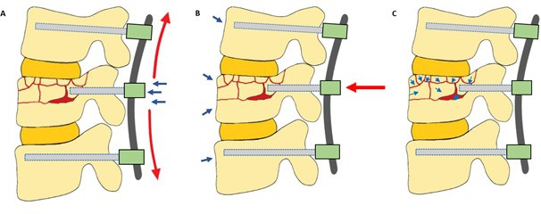
Objective: In the present study, the authors our technique utilizing unipedicular index vertebra fixation and manipulation in MIS for thoracolumbar fractures with burst elements. To our knowledge, this technique is not well described in literature as open approaches are often adopted for the above. The authors sought to highlight the 2-year radiological and functional outcomes of 20 consecutive patients who underwent this technique.
Methods: A retrospective review of prospectively collected data was conducted on 20 patients with thoracolumbar fractures with burst elements who underwent fixation using our technique. Patient data collected included demographic characteristics, mechanism of injury, associated injuries, neurological deficit at the time of admission, pre- and postoperative neurological evaluation, and length of hospital stay. Radiological investigations included plain radiographs, computed tomography of the spine with reconstruction, and magnetic resonance imaging of the spine, which provided data for radiological fracture classifications such as AO Spine and derivation of Thoracolumbar Injury Classification and Severity Score, as well as preoperative planning. Radiological investigations in the postoperative period were carried out by standing radiographs or EOS whole spine at each postoperative follow-up for up to 2 years. Radiological parameters-vertebral wedge angle, regional kyphosis angle, coronal Cobb angle, and anterior and posterior vertebral body heights-were recorded at preoperative, intraoperative, postoperative, and up to 2-year follow-up. Clinical outcome scores (visual analog score [VAS] and Oswestry Disability Index [ODI]) were also recorded at similar timepoints.
Results: Radiological outcomes reflect significant lordotic corrections of the vertebral wedge angles up to 2-year follow-up when compared with preoperative values (intraoperative: P = 0.06; postoperative: P = 0.001; 3 months: P = 0.002; 6 months: P = 0.004; 1 year: P = 0.011; 2 years: P = 0.016). Additionally, significant lordotic corrections of regional kyphosis angles (intraoperative: P = 0.00; postoperative: P = 0.00; 3 months: P = 0.031; 6 months: P = 0.039) and increases in anterior vertebral body heights (postoperative: P = 0.001; 3 months: P = 0.010; 6 months: P = 0.020) at up to 6-month follow-up were found. Preoperatively, median VAS of 85 (range 30-100) and ODI of 90 (range 40-98) were recorded. Statistically significant improvements in VAS and ODI were found across all timepoints when compared with preoperative values, with a mean VAS of 11.5 (SD 4.8) and ODI of 9.9 (SD 4.5) at 2-year follow-up.
Conclusion: Surgical management of thoracolumbar fractures with or without neurological deficit has a role in reducing nursing requirements and postoperative morbidity in patients with polytrauma and other associated injuries. Our approach in treating thoracolumbar fractures with burst elements using MIS short-segment fixation and unipedicular screw manipulation technique shows satisfactory radiological correction and high rates of fracture union while reducing approach-related morbidity and improving functional outcomes.
Keywords: burst fracture; flexion-distraction, indirect reduction; minimally invasive surgery; short-segment instrumentation; thoracolumbar.
This manuscript is generously published free of charge by ISASS, the International Society for the Advancement of Spine Surgery. Copyright © 2023 ISASS. To see more or order reprints or permissions, see http://ijssurgery.com.
Conflict of interest statement
Declaration of Conflicting Interests : The authors report no conflicts of interest in this work.
Figures


References
-
- Takami M, Yamada H, Nohda K, Yoshida M. A minimally invasive surgery combining temporary percutaneous pedicle screw fixation without fusion and vertebroplasty with transpedicular intracorporeal hydroxyapatite blocks grafting for fresh thoracolumbar burst fractures: prospective study. Eur J Orthop Surg Traumatol. 2014;24 Suppl 1:S159–S165. 10.1007/s00590-013-1266-2 - DOI - PubMed
LinkOut - more resources
Full Text Sources