. 2023 Jul 24;13(1):11899.
doi: 10.1038/s41598-023-38659-9.

Anandamide modulates WNT-5A/BCL-2, IP3/NFATc1, and HMGB1/NF-κB trajectories to protect against mercuric chloride-induced acute kidney injury

Affiliations

Anandamide modulates WNT-5A/BCL-2, IP3/NFATc1, and HMGB1/NF-κB trajectories to protect against mercuric chloride-induced acute kidney injury

Dalaal M Abdallah et al. Sci Rep. .

Abstract

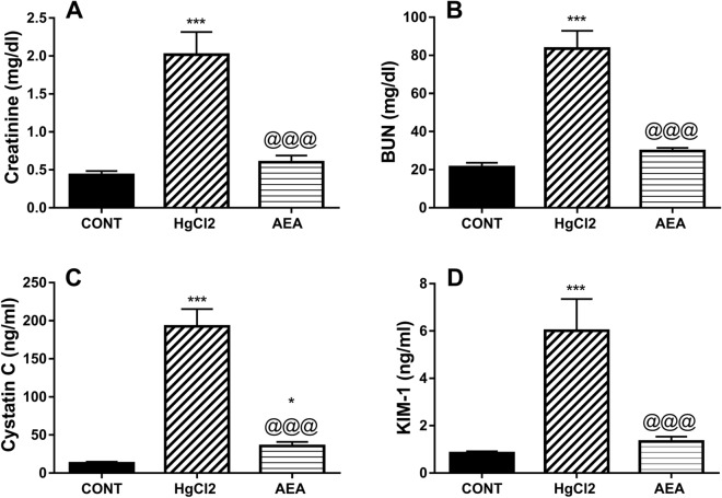
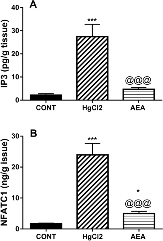
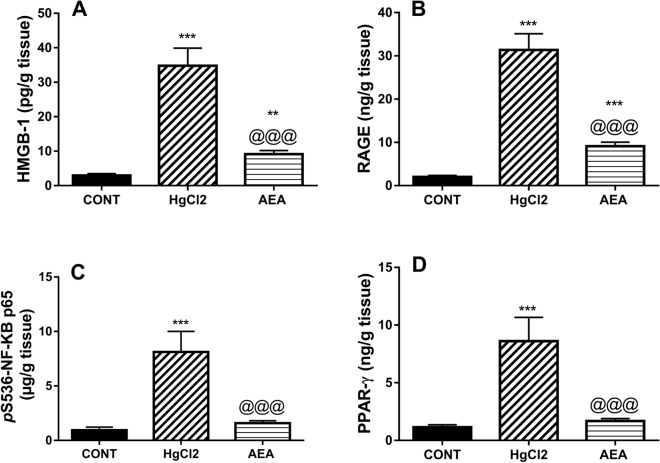
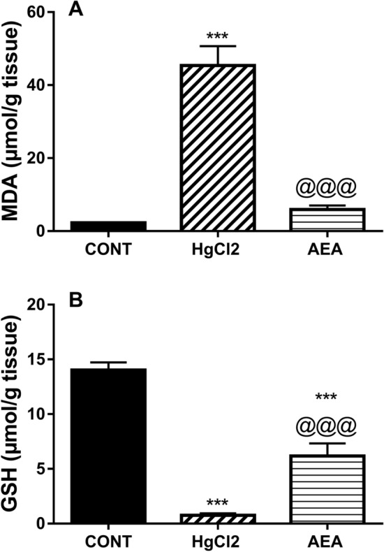
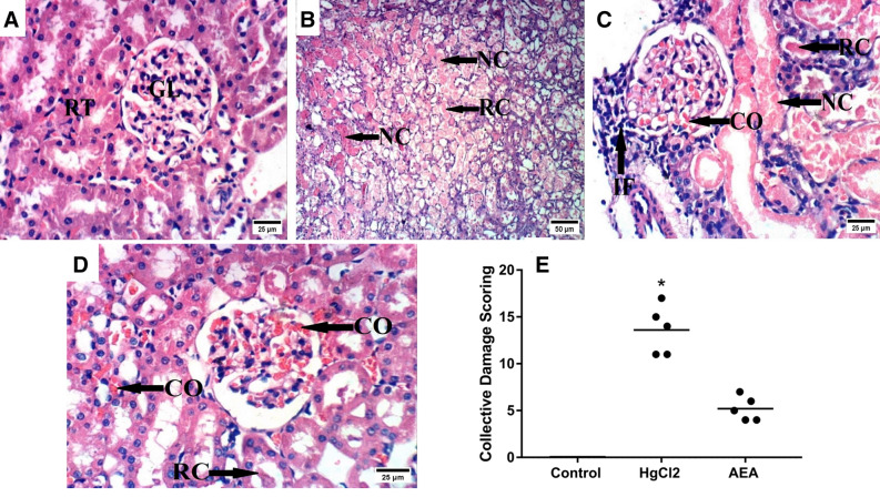
Endocannabinoid anandamide (AEA) has a physiological role in regulating renal blood flow, whereas its analogs ameliorated renal ischemia/reperfusion injury. Nonetheless, the role of AEA against mercuric chloride (HgCl2)-induced renal toxicity has not been unraveled. Rats were allocated into control, HgCl2, and HgCl2/AEA treated groups. The administration of AEA quelled the HgCl2-mediated increase in inositol trisphosphate (IP3) and nuclear factor of activated T-cells cytoplasmic 1 (NFATc1). The endocannabinoid also signified its anti-inflammatory potential by turning off the inflammatory cascade evidenced by the suppression of high mobility group box protein-1 (HMGB1), receptor of glycated end products (RAGE), nuclear factor-κB p65 (NF-κB), and unexpectedly PPAR-γ. Additionally, the aptitude of AEA to inhibit malondialdehyde and boost glutathione points to its antioxidant capacity. Moreover, AEA by enhancing the depleted renal WNT-5A and reducing cystatin-C and KIM-1 (two kidney function parameters) partly verified its anti-apoptotic capacity, confirmed by inhibiting caspase-3 and increasing B-cell lymphoma-2 (BCL-2). The beneficial effect of AEA was mirrored by the improved architecture and kidney function evidenced by the reduction in cystatin-C, KIM-1, creatinine, BUN, and caspase1-induced activated IL-18. In conclusion, our results verify the reno-protective potential of AEA against HgCl2-induced kidney injury by its anti-inflammatory, antioxidant, and anti-apoptotic capacities by modulating WNT-5A/BCL-2, IP3/NFATC1, HMGB-1/RAGE/NF-κB, caspase-1/IL-18, and caspase-3/BCL-2 cues.


Conflict of interest statement

The authors declare no competing interests.

Figures

Figure 1
Figure 1
Effect of AEA on serum (A) creatinine, (B) BUN, (C) cystatin-C, and (D) KIM-1 in HgCl2-induced AKI in rats. Data are expressed as the mean of 6 rats ± SD. *P < 0.05 and ***P < 0.001 compared with CONT and @@@P < 0.001 compared with HgCl2 groups using one-way ANOVA followed by Tukey’s Multiple Comparison post hoc test. AEA anandamide, AKI acute kidney injury, BUN blood urea nitrogen, CONT control, HgCl2 mercuric chloride, KIM-1 kidney injury molecule-1.
Figure 2
Figure 2
Effect of AEA on renal contents of (A) IP3 and (B) NFATc1 in HgCl2-induced AKI in rats. Data are expressed as the mean of 6 rats ± SD. *P < 0.05 and ***P < 0.001 compared with CONT and @@@P < 0.001 compared with HgCl2 groups using one-way ANOVA followed by Tukey's Multiple Comparison post hoc test. AEA anandamide, AKI acute kidney injury, CONT control, HgCl2 mercuric chloride, IP3 inositol trisphosphate, NFATc1 nuclear factor of activated T-cells cytoplasmic-1.
Figure 3
Figure 3
Effect of AEA on renal contents of (A) HMGB-1, (B) RAGE, (C) pS536-NF-κB p65, and (D) PPAR-γ in HgCl2-induced AKI in rats. Data are expressed as the mean of 6 rats ± SD. **P < 0.01 and ***P < 0.001 compared with CONT and @@@P < 0.001 compared with HgCl2 groups using one-way ANOVA followed by Tukey's Multiple Comparison post hoc test. AEA anandamide, AKI acute kidney injury, CONT control, HgCl2 mercuric chloride, HMGB-1 high mobility group box, pS536-NF-κB p65 nuclear factor-κB p65 phosphorylated at serine 536, PPAR-γ peroxisome proliferator-activated receptor gamma, RAGE receptor for advanced glycation end products.
Figure 4
Figure 4
Effect of AEA on renal content of (A) caspase-1 and serum level (B) IL-18 in HgCl2-induced AKI in rats. Data are expressed as the mean of 6 rats ± SD. **P < 0.01 and ***P < 0.001 compared with CONT and @@@P < 0.001 compared with HgCl2 groups using one-way ANOVA followed by Tukey's Multiple Comparison post hoc test. AEA anandamide, AKI acute kidney injury, CONT control, HgCl2 mercuric chloride, IL-18 interleukin 18.
Figure 5
Figure 5
Effect of AEA on renal contents of (A) MDA and (B) GSH in HgCl2-induced AKI in rats. Data are expressed as the mean of 6 rats ± SD. ***P < 0.001 compared with CONT and @@@P < 0.001 compared with HgCl2 groups using one-way ANOVA followed by Tukey's Multiple Comparison post hoc test. AEA anandamide, AKI acute kidney injury, CONT control, GSH glutathione, HgCl2 mercuric chloride, MDA malondialdehyde.
Figure 6
Figure 6
Effect of AEA on renal contents of (A) WNT-5A and (B) BCL-2, as well as (C) caspase-3 activity in HgCl2-induced AKI in rats. Data are expressed as the mean of 6 rats ± SD. **P < 0.01 and ***P < 0.001 compared with CONT and @@@P < 0.001 compared with HgCl2 groups using one-way ANOVA followed by Tukey's Multiple Comparison post hoc test. AEA anandamide, AKI acute kidney injury, BCL-2 B-cell lymphoma-2, CONT control, HgCl2 mercuric chloride.
Figure 7
Figure 7
Effect of AEA on HgCl2-induced histopathology alterations. Microscopically, the section of the (A) normal control group reveals the normal histological architecture of renal parenchyma (normal renal tubules and glomeruli). In contrast, the sections of the (B,C) HgCl2-untreated rats show notable histopathological damage characterized by congestion of glomerular tufts, marked coagulative necrosis of epithelial lining renal tubules, renal cast in the lumen of renal tubules, and inflammatory cells infiltration. On the other hand, the section of the (D) AEA-treated group shows an improved picture with milder congestion of glomerular tufts and renal blood vessels as well as renal cast in the lumen of some renal tubules. All black arrows point to the altered structure in the section (scale bar = 25;50). Panel (E) illustrates the collective kidney damage score scatter blot; and data were analyzed using the Kruskal–Wallis test followed by Dunn’s post hoc test. Values were calculated from 5 fields of n = 3 rats/group; as compared with *P < 0.05 compared with CONT group. AEA anandamide, HgCl2 mercuric chloride.

Similar articles

Cited by

References

    1. Park JD, Zheng W. Human exposure and health effects of inorganic and elemental mercury. J. Prev. Med. Public Health. 2012;45:344–352. doi: 10.3961/jpmph.2012.45.6.344. - DOI - PMC - PubMed
    1. Doering S, Bose-O’Reilly S, Berger U. Essential indicators identifying chronic inorganic mercury intoxication: Pooled analysis across multiple cross-sectional studies. PLoS One. 2016;11:e0160323. doi: 10.1371/journal.pone.0160323. - DOI - PMC - PubMed
    1. Zalups RK. Molecular interactions with mercury in the kidney. Pharmacol. Rev. 2000;52:113–143. - PubMed
    1. Ajsuvakova OP, et al. Sulfhydryl groups as targets of mercury toxicity. Coord. Chem. Rev. 2020;417:213343. doi: 10.1016/j.ccr.2020.213343. - DOI - PMC - PubMed
    1. Yeh JH, Chung HM, Ho CM, Jan CR. Mercury-induced Ca2+ increase and cytotoxicity in renal tubular cells. Life Sci. 2004;74:2075–2083. doi: 10.1016/j.lfs.2003.09.046. - DOI - PubMed

Publication types