. 2023 Jul 24;13(1):11907.
doi: 10.1038/s41598-023-39102-9.

Molecular features driving condensate formation and gene expression by the BRD4-NUT fusion oncoprotein are overlapping but distinct

Affiliations

Molecular features driving condensate formation and gene expression by the BRD4-NUT fusion oncoprotein are overlapping but distinct

Martyna Kosno et al. Sci Rep. .

Abstract

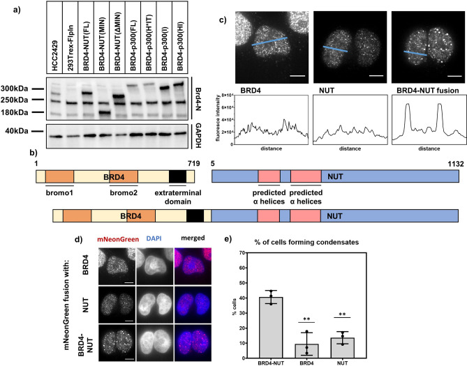
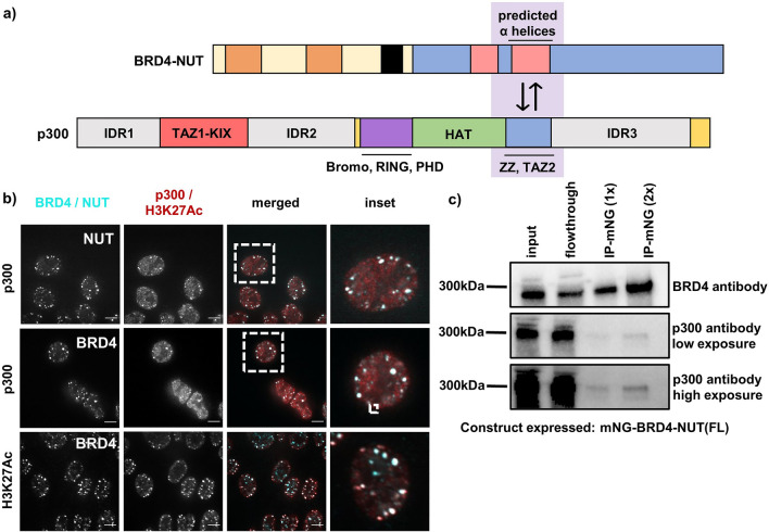
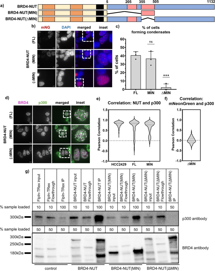
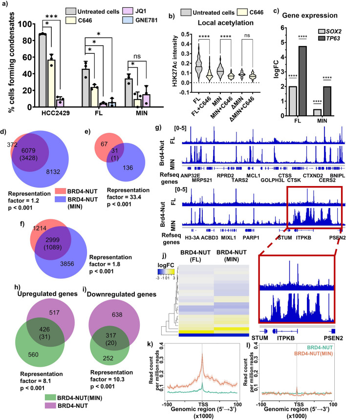
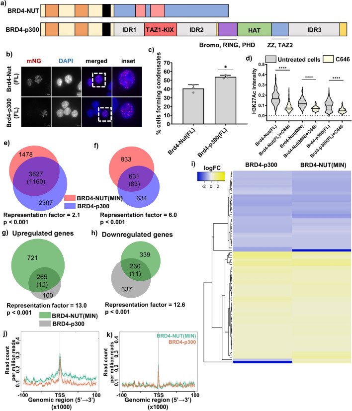
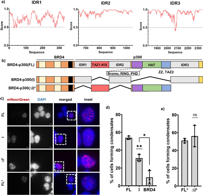
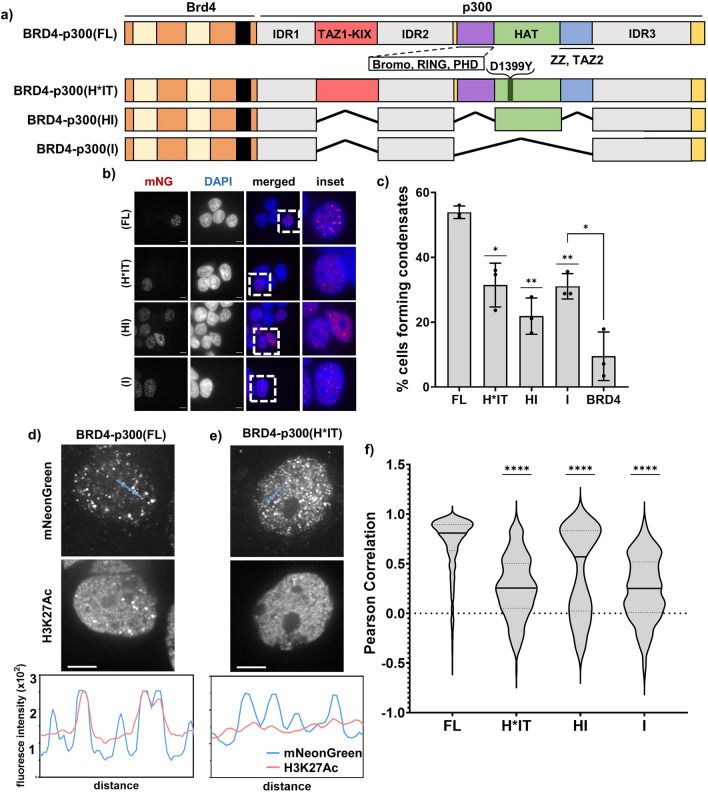
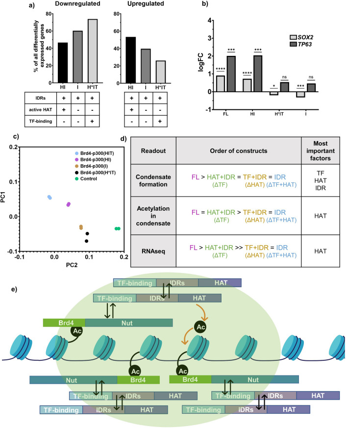
Aberrant formation of biomolecular condensates has been proposed to play a role in several cancers. The oncogenic fusion protein BRD4-NUT forms condensates and drives changes in gene expression in Nut Carcinoma. Here we sought to understand the molecular elements of BRD4-NUT and its associated histone acetyltransferase (HAT), p300, that promote these activities. We determined that a minimal fragment of NUT (MIN) in fusion with BRD4 is necessary and sufficient to bind p300 and form condensates. Furthermore, a BRD4-p300 fusion protein also forms condensates and drives gene expression similarly to BRD4-NUT(MIN), suggesting the p300 fusion may mimic certain features of BRD4-NUT. The intrinsically disordered regions, transcription factor-binding domains, and HAT activity of p300 all collectively contribute to condensate formation by BRD4-p300, suggesting that these elements might contribute to condensate formation by BRD4-NUT. Conversely, only the HAT activity of BRD4-p300 appears necessary to mimic the transcriptional profile of cells expressing BRD4-NUT. Our results suggest a model for condensate formation by the BRD4-NUT:p300 complex involving a combination of positive feedback and phase separation, and show that multiple overlapping, yet distinct, regions of p300 contribute to condensate formation and transcriptional regulation.


Conflict of interest statement

The authors declare no competing interests.

Figures

Figure 1
Figure 1
BRD4-NUT fusion protein forms nuclear condensates in human cells. (a) Western blot showing the expression levels between stable cell lines expressing different constructs. All cell lines treated with 5 ng/mL Doxycycline for 2 h, followed by 4 h washout. Antibodies used include BRD4(N) and GAPDH as a loading control. (b) Cartoon of BRD4, NUT and BRD4-NUT fusion proteins. Bromodomains 1 and 2 and extraterminal domain of BRD4 as well as predicted α-helices are indicated. (c) Line profiles across cells expressing mNeonGreen-tagged BRD4, NUT and BRD4-NUT fusion; scale bars = 10 μm. (d) Micrographs of cells expressing mNeonGreen-tagged BRD4, NUT and BRD4-NUT; scale bars = 10 μm. (e) Quantification of percentage of cells forming condensates (at least 2 condensates larger than 1.25 μm in diameter).
Figure 2
Figure 2
BRD4-NUT condensates recruit p300 histone acetyltransferase and are heavily acetylated at H3K27. (a) Cartoon of BRD4-NUT and p300 interaction; known interaction motifs are indicated. (b) Micrographs of HCC2429 Nut Carcinoma cells co-stained with BRD4 and p300 or BRD4 and Histone H3K27Ac antibodies; scale bar = 10 μm. (c) Immunoprecipitation against mNeonGreen; western blot with a p300 antibody showing that BRD4-NUT pulls down p300.
Figure 3
Figure 3
Minimal p300-interaction fragment of NUT in BRD4-NUT fusion is necessary and sufficient for condensate formation. (a) Schematic of all BRD4-NUT constructs. (b) Representative micrographs showing condensate formation in stable cell lines expressing different constructs. Staining against mNeonGreen shown in red, overlay with DAPI to indicate the nucleus. Scale bars = 10 μm. (c) Quantification of the micrographs represented in (b). (d) Representative micrographs comparing expression of constructs with or without p300-interaction motif in cells. Cells stained with anti-BRD4 antibody (magenta) and anti-p300 antibody (green). Scale bars = 10 μm. (e) and (f) Quantification of the overlap between condensates via co-staining shown as Pearson correlation. Cells co-stained with NUT and p300 antibodies (left) or mNeonGreen and p300 antibodies (right); Nut antibody epitope was removed in BRD4-NUT(ΔMIN), see Fig. S2b for more details. (g) Immunoprecipitation against mNeonGreen; western blot with p300 antibody and BRD4(N) antibody.
Figure 4
Figure 4
p300 binding and enzymatic activity are necessary for gene expression changes induced by BRD4-NUT. (a) Quantification of cells capacity to form condensates, comparing cells that are untreated (gray) and cells treated for 1 h with 20 µM C646 inhibitor (yellow), for 1 h with 1 µM JQ1 inhibitor (pink) or for 3 h with 1 µM GNE-781 inhibitor (blue). Cells compared here include HCC2429 Nut Carcinoma cell line and stable cell lines expressing BRD4-NUT(FL) and BRD4-NUT(MIN). Representative images of cells treated with inhibitors shown in Fig. S8. (b) Local acetylation measured as average fluorescence intensity across a condensate. Data shown for cells untreated (gray) or treated with C646 (yellow), in cells expressing BRD4-NUT(FL), BRD4-NUT(MIN) and BRD4-NUT(ΔMIN). (c) Graph showing logFC for SOX2 and TP63 genes upon expression of different BRD4-p300 constructs. (d) Venn diagram of gene occupancy by BRD4-NUT(FL) (pink) and BRD4-NUT(MIN) (blue), found via ChIPseq with α-NUT antibody: all annotated genes occupied by either protein. Values in the diagram show the number of unique genes annotated; numbers in parentheses represent the mean overlap in 20 iterations with a randomly generated gene pool of the same size, from the human genome. (e) Venn diagram of gene occupancy by BRD4-NUT(FL) (pink) and BRD4-NUT(MIN) (blue), found via ChIPseq with α-NUT antibody: genes annotated at promoter-TSS regions of genes, with normalized signal value of 0.3–1. Values in the diagram as in (d). (f) Venn diagram of gene occupancy by BRD4-NUT(FL) (pink) and BRD4-NUT(MIN) (blue), found via ChIPseq with α-NUT antibody: genes occupied by either protein outside of the promoter-TSS region, within 500 kb of TSS. Values in the diagram as in (d). (g) Example ChIPseq tracks comparing gene occupancy by BRD4-NUT(FL) and BRD4-NUT(MIN). Data scale: 0–5. Inset shows more closely an example of the same genes being occupied by both proteins, but BRD4-NUT(MIN) occupying more loci. (h) Venn diagram of genes upregulated upon expression of BRD4-NUT(FL) (purple) or BRD4-NUT(MIN) (green), found via RNAseq. Genes shown when fold change in expression was greater than two and p < 0.05. Numbers in parentheses represent the mean overlap in 20 iterations with a randomly generated gene pool of the same size, from the human genome. (i) Venn diagram of genes downregulated upon expression of BRD4-NUT(FL) (purple) or BRD4-NUT(MIN) (green), found via RNAseq. Statistical analyses as in (g). (j) RNAseq-ChIPseq data integration: heatmap showing up- and down-regulated genes found via RNAseq, bound by both BRD4-NUT(FL) and BRD4-NUT(MIN). Top 200 genes shown in the heatmap, based on p value. (k) Profiles of enrichment of BRD4-NUT (green) and BRD4-NUT(MIN) (orange) of differentially upregulated genes within 100 kb from TSS. Legend same as in (l). (l) Profiles of enrichment of BRD4-NUT (green) and BRD4-NUT(MIN) (orange) of differentially downregulated genes within 100 kb from TSS.
Figure 5
Figure 5
BRD4-p300 fusion forms condensates and recapitulates many of the transcriptional changes observed for BRD4-NUT fusion. (a) Schematic of BRD4-NUT and BRD4-p300. (b) Micrographs of BRD4-NUT and BRD4-p300—expressing stable cell lines. Cells are stained with mNeonGreen antibody; scale bars = 10 μm. (c) Quantification of the micrographs represented in (b). (d) Quantification of histone H3K27Ac staining in untreated vs. C646-treated cells. Local acetylation shown as average fluorescence intensity per condensate. (e) Venn diagram of genes acetylated upon expression of BRD4-NUT(MIN) (pink) and BRD4-p300 (blue), found via ChIPseq with α-H3K27Ac antibody, at promoter-TSS regions: all annotated genes. Values in the diagram show the number of unique genes annotated; numbers in parentheses represent the mean overlap in 20 iterations with a randomly generated gene pool of the same size, from the human genome. (f) Venn diagram of genes acetylated upon expression of BRD4-NUT(MIN) (pink) and BRD4-p300 (blue), found via ChIPseq with α-H3K27Ac antibody, at promoter-TSS regions: Diagram shows only genes after applying 0.3–1 normalized signal value cutoff. Values in the diagram as in (e). (g) Venn diagram of genes upregulated upon expression of BRD4-NUT(MIN) (green) or BRD4-p300 (gray), found via RNAseq. Genes shown when fold change in expression was greater than two and p < 0.05. Numbers in parentheses represent the mean overlap in 20 iterations with a randomly generated gene pool of the same size, from the human genome. (h) Venn diagram of genes downregulated upon expression of BRD4-NUT(MIN) (green) or BRD4-p300 (gray), found via RNAseq. Statistical analyses as in (g). (i) RNAseq-ChIPseq data integration: heatmap showing up- and down-regulated genes found via RNAseq, that are bound by both BRD4-NUT(MIN) and BRD4-p300. Top up- and downregulated genes shown in the heatmap, based on log fold change, where log fold change was greater than 0.5. (j) Profiles of enrichment of BRD4-NUT(MIN) (green) and BRD4-p300 (orange) of differentially upregulated genes within 100 kb from TSS. Legend same as in (k). (k) Profiles of enrichment of BRD4-NUT(MIN) (green) and BRD4-p300 (orange) of differentially downregulated genes within 100 kb from TSS.
Figure 6
Figure 6
p300 IDRs are dispensable for condensate formation. (a) Disorder prediction of p300 IDRs as shown via IUPRED2 analysis. (b) Schematics of BRD4-p300 (FL), (IDR) and (ΔIDR) constructs; different domains and disordered regions are indicated. (c) Micrographs of all the BRD4-p300 constructs from (a); scale bars = 10 µm. Constructs marked with an asterisk were expressed via transient transfection. (d) Quantification of the micrographs represented in (c), expressed in stable cell lines. (e) Quantification of the micrographs represented in (c), expressed via transient transfection.
Figure 7
Figure 7
IDRs, HAT and TF binding domains of p300 collectively contribute to condensate formation. (a) Schematic of BRD4-p300—based constructs, including FL, H*IT, HI and I. (b) Micrographs of all the BRD4-p300 constructs from (a); scale bars = 10 μm. (c) Quantification of the micrographs represented in (b). (d) Colocalization of BRD4-p300(FL), as visualized by α-mNeonGreen staining, with α-H3K27Ac staining, shown in micrographs and via line profiles. (e) Colocalization of BRD4-p300(H*IT), as visualized by α-mNeonGreen staining, with α-H3K27Ac staining, shown in micrographs and via line profiles. (f) Quantification of correlation between α-mNeonGreen and α-H3K27Ac condensates, as shown via Pearson Correlation.
Figure 8
Figure 8
Acetylation activity of p300 is necessary for transcriptional changes observed upon expression of BRD4-p300(FL). (a) Percent of genes affected upon expression of different BRD4-p300 constructs. (b) Graph showing logFC for SOX2 and TP63 genes upon expression of different BRD4-p300 constructs. (c) PCA plot, summarizing RNAseq data for cells expressing all constructs. Cell lines excluded in this plot: BRD4-NUT(FL) and BRD4-NUT(MIN) are included in the full PCA in Fig. S4b. (d) Summary of molecular parts of p300 required for condensate formation, condensate-localized acetylation and transcriptional changes in cells. (e) Final model of condensate formation.

Update of

Similar articles

Cited by

References

    1. Shirnekhi HK, Chandra B, Kriwacki RW. The role of phase-separated condensates in fusion oncoprotein-driven cancers. Ann. Rev. Cancer Biol. 2023;7:73–91. doi: 10.1146/annurev-cancerbio-061421-122050. - DOI
    1. Chandra B, et al. Phase separation mediates NUP98 fusion oncoprotein leukemic transformation. Cancer Discov. 2022;12(4):1152–1169. doi: 10.1158/2159-8290.CD-21-0674. - DOI - PMC - PubMed
    1. Terlecki-Zaniewicz S, et al. Biomolecular condensation of NUP98 fusion proteins drives leukemogenic gene expression. Nat. Struct. Mol. Biol. 2021;28(2):190–201. doi: 10.1038/s41594-020-00550-w. - DOI - PMC - PubMed
    1. Boulay G, et al. Cancer-specific retargeting of BAF complexes by a prion-like domain. Cell. 2017;171(1):163–178. doi: 10.1016/j.cell.2017.07.036. - DOI - PMC - PubMed
    1. Qin Z, et al. Phase separation of EML4-ALK in firing downstream signaling and promoting lung tumorigenesis. Cell Discov. 2021;7(1):33. doi: 10.1038/s41421-021-00270-5. - DOI - PMC - PubMed

Publication types