. 2023 Jul 24;13(1):11930.
doi: 10.1038/s41598-023-39033-5.

Acoustic and optoacoustic stimulations in auditory brainstem response test in salicylate induced tinnitus

Affiliations

Acoustic and optoacoustic stimulations in auditory brainstem response test in salicylate induced tinnitus

Katayoon Montazeri et al. Sci Rep. .

Abstract

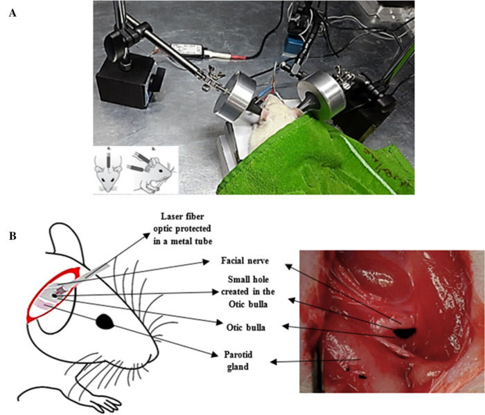
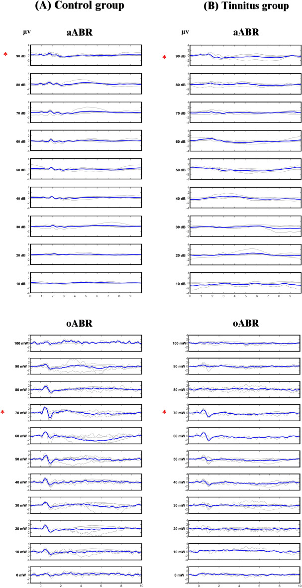
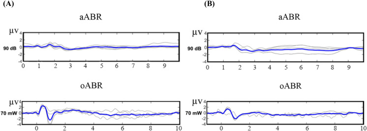
As a common debilitating disorder worldwide, tinnitus requires objective assessment. In the auditory brainstem response (ABR) test, auditory potentials can be evoked by acoustic or optoacoustic (induced by laser light) stimulations. In order to use the ABR test in the objective assessment of tinnitus, in this study, acoustic ABR (aABR) and optoacoustic ABR (oABR) were compared in the control and tinnitus groups to determine the changes caused by sodium salicylate (SS)-induced tinnitus in rat. In both aABR and oABR, wave II was the most prominent waveform, and the amplitude of wave II evoked by oABR was significantly higher than that of aABR. Brainstem transmission time (BTT), which represents the time required for a neural stimulation to progress from the auditory nerve ending to the inferior colliculus, was significantly shorter in oABR. In the tinnitus group, there was a significant increase in the threshold of both ABRs and a significant decrease in the amplitude of wave II only in the oABR. Based on our findings, the ABR test has the potential to be used in the assessment of SS-induced tinnitus, but oABR has the advantages of producing more prominent waveforms and significantly reducing the amplitude of wave II in tinnitus.


Conflict of interest statement

The authors declare no competing interests.

Figures

Figure 1
Figure 1
Electrophysiological tests of aABR and oABR using acoustic and optoacoustic stimulations. (A) Three surface electrodes were placed on the scalp of the anesthetized animal to record the aABR by the audiology lab device. (B) Ear surgery was performed to access the cochlea and laser irradiation for oABR test. The Otic bulla was exposed through a posterior tympanum approach. The facial nerve and parotid gland were the landmarks for finding the Otic bulla, where a small hole was created to expose the cochlea. The Audiology Lab device was synchronized with the laser device to record the evoked potentials immediately after the laser irradiation. The laser fiber optic, which was placed in a metal tube for protection, was fixed by a mechanical holder in a non-contact mode at a distance of 1 mm from the hole created in the Otic bulla in the direction of the cochlea.
Figure 2
Figure 2
aABR and oABR waveforms. (A) Control group, (B) tinnitus group. In each row, the bold line represents the grand average of the waveforms. The highest mean amplitude of wave II, which was the most prominent waveform in both aABR and oABR, occurred at 90 dB SPL aABR and 70 mW oABR power, indicated by *.
Figure 3
Figure 3
Single waveforms of aABR at 90 dB SPL and oABR at 70 mW from control and tinnitus groups were set side by side for comparison. The bold line in each group represents the grand average. (A) Control group, (B) Tinnitus group.
Figure 4
Figure 4
Flow diagram of method and study design. GPIAS gap prepulse inhibition of acoustic startle, GIN gap-in-noise, PPI pre-pulse inhibition, aABR acoustic auditory brainstem response, oABR optoacoustic auditory brainstem response, SS sodium salicylate, mg milligram, kg kilogram.

References

    1. Baguley D, McFerran D, Hall D. Tinnitus. The Lancet. 2013;382:1600–1607. doi: 10.1016/S0140-6736(13)60142-7. - DOI - PubMed
    1. Jarach CM, et al. Global prevalence and incidence of tinnitus: A systematic review and meta-analysis. JAMA Neurol. 2022;79:888–900. doi: 10.1001/jamaneurol.2022.2189. - DOI - PMC - PubMed
    1. Hullfish J, Abenes I, Yoo HB, De Ridder D, Vanneste S. Frontostriatal network dysfunction as a domain-general mechanism underlying phantom perception. Hum. Brain Mapp. 2019;40:2241–2251. doi: 10.1002/hbm.24521. - DOI - PMC - PubMed
    1. Langguth B, Kreuzer PM, Kleinjung T, De Ridder D. Tinnitus: Causes and clinical management. Lancet Neurol. 2013;12:920–930. doi: 10.1016/S1474-4422(13)70160-1. - DOI - PubMed
    1. Tang D, Li H, Chen L. Advances in understanding, diagnosis, and treatment of tinnitus. Adv. Exp. Med. Biol. 2019;1130:109–128. doi: 10.1007/978-981-13-6123-4_7. - DOI - PubMed

Publication types