

Nucleus high intensity in the T2-weighted MRI is a potential predictor of annulus tear in cervical injured patients: a case comparative study
- PMID: 37488519
- PMCID: PMC10364398
- DOI: 10.1186/s12891-023-06615-3
Nucleus high intensity in the T2-weighted MRI is a potential predictor of annulus tear in cervical injured patients: a case comparative study
Abstract
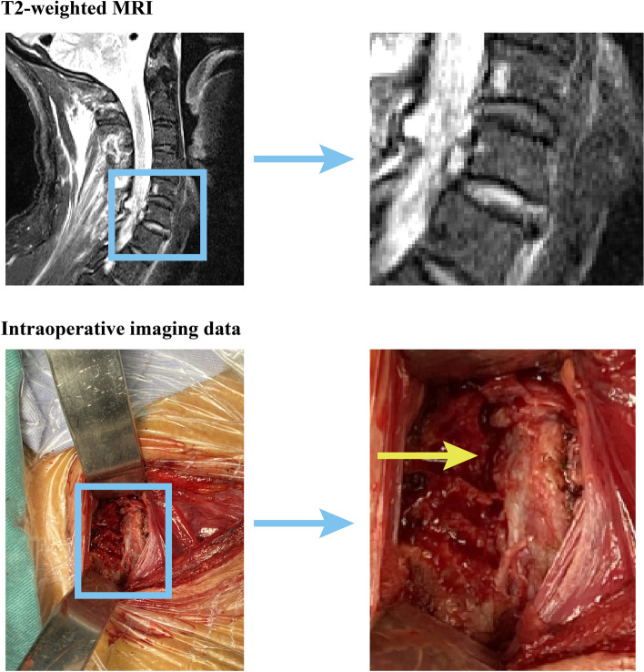
Background: Segmental fusion operations assume paramount significance for individuals afflicted by full layers of annulus tears as they avert the perils of rapid disc degeneration and segmental instability. Structures with high signal intensity in the T2-weighted MRI can predict potential damage to the injured segment. Since local structures are shortly related biomechanically, this may be an effective predictor for annulus tears.
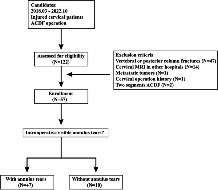
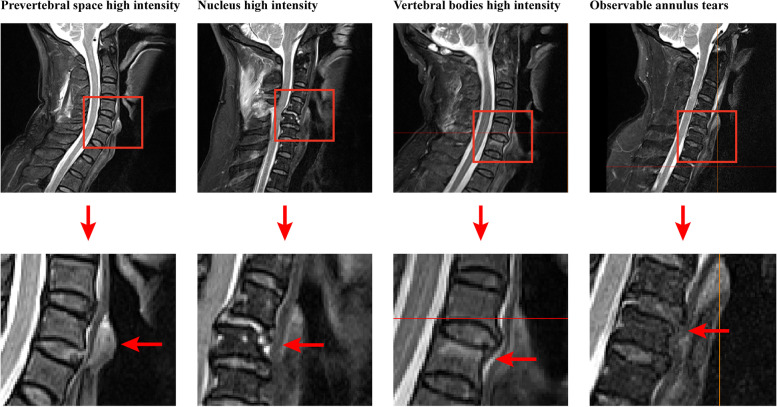
Methods: A retrospective analysis of the clinical data of 57 patients afflicted by cervical injuries and subjected to single-segment ACDF has been performed in this study. The surgeon performed intraoperative exploration to assess the integration status of the annulus. The signal intensity of the prevertebral space, nucleus, and injured vertebral bodies were judged in the T2-weighted imaging data. Regression analyses identified independent predictors for annulus tears, and the area under the receiver operating characteristic curve (AUC) was computed to evaluate the predictive performance of potential independent predictors.
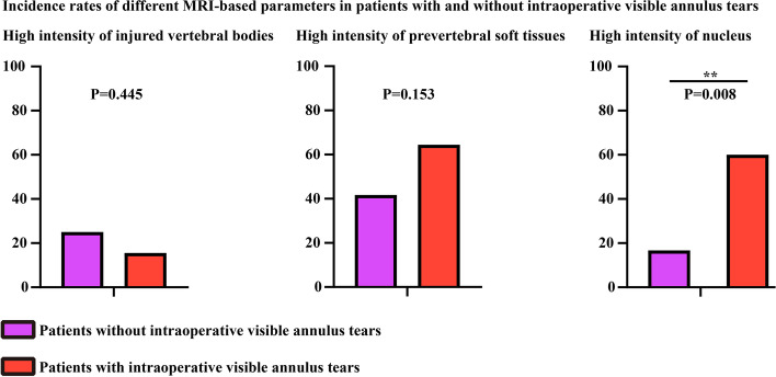
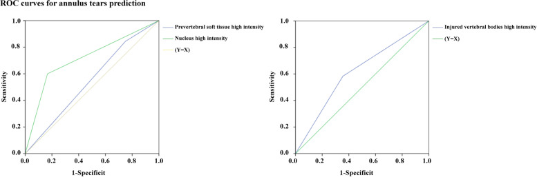
Results: The occurrence of nucleus high intensity was significantly higher among individuals with annulus tears, and the nucleus high intensity was deemed an independent predictor for determining the presence of intraoperative visible annulus tears in patients with cervical injuries. AUC for nucleus high intensity was calculated as 0.717, with a corresponding p-value less than 0.05.
Conclusions: In the realm of diagnosing annulus tears in injured cervical patients, nucleus high intensity in the T2-weighted MRI emerges as a promising predictive factor. Notably, this applies specifically to patients devoid of fracture and visible annulus tears in their MRI scans. Such positive outcomes should be regarded as prospective indications for ACDF.
Keywords: Annulus tears; Independent predictors; Injured cervical patients; Nucleus high intensity; surgicalSurgical indications.
© 2023. The Author(s).
Conflict of interest statement
The authors declare no competing interests.
Figures
Similar articles
-
Total disc replacement using a tissue-engineered intervertebral disc in vivo: new animal model and initial results.Evid Based Spine Care J. 2010 Aug;1(2):62-6. doi: 10.1055/s-0028-1100918. Evid Based Spine Care J. 2010. PMID: 23637671 Free PMC article.
-
Disc degeneration assessed by quantitative T2* (T2 star) correlated with functional lumbar mechanics.Spine (Phila Pa 1976). 2013 Nov 15;38(24):E1533-40. doi: 10.1097/BRS.0b013e3182a59453. Spine (Phila Pa 1976). 2013. PMID: 23921323 Free PMC article.
-
Is there an impact of cervical plating on the development of adjacent segment degeneration following Smith-Robinson procedure? A magnetic resonance imaging study of 84 patients with a 24-year follow-up.Spine J. 2019 Apr;19(4):587-596. doi: 10.1016/j.spinee.2018.09.001. Epub 2018 Sep 6. Spine J. 2019. PMID: 30195935
-
Cervical intervertebral disc calcification with extreme lateral herniation in a child: T2-weighted signal intensity of the involved disc can be restored to normal.Childs Nerv Syst. 2016 Apr;32(4):749-52. doi: 10.1007/s00381-015-2886-0. Epub 2015 Aug 29. Childs Nerv Syst. 2016. PMID: 26319586
-
Intervertebral disc degeneration.Eur Spine J. 1993 Mar;1(4):205-13. doi: 10.1007/BF00298361. Eur Spine J. 1993. PMID: 20054919 Review.
References
-
- Haughton VM, Schmidt TA, Keele K, An HS, Lim TH. Flexibility of lumbar spinal motion segments correlated to type of tears in the annulus fibrosus. J Neurosurg. 2000;92:81–86. - PubMed
MeSH terms
LinkOut - more resources
Full Text Sources
Medical
