Case Reports
. 2023 Jul 25;17(1):317.
doi: 10.1186/s13256-023-04048-z.

Pathogenesis of spinal intramedullary lipomas: two case reports

Affiliations
Case Reports

Pathogenesis of spinal intramedullary lipomas: two case reports

Luis Miguel Moreno Gómez et al. J Med Case Rep. .

Abstract

Background: Spinal lipomas not associated with dysraphism are rare and have an unknown natural history. In this report, we describe two cases; they showed recurrence during long-term follow-up, which makes us doubt a benign malformative etiology.

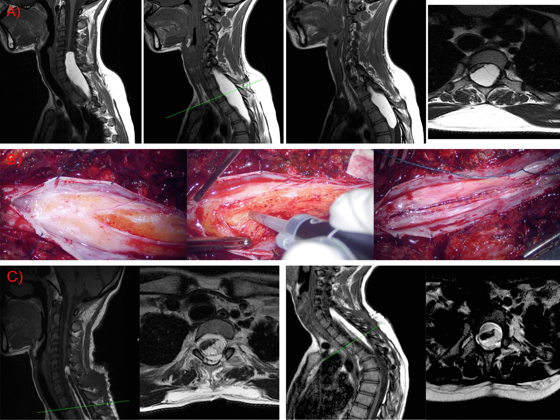
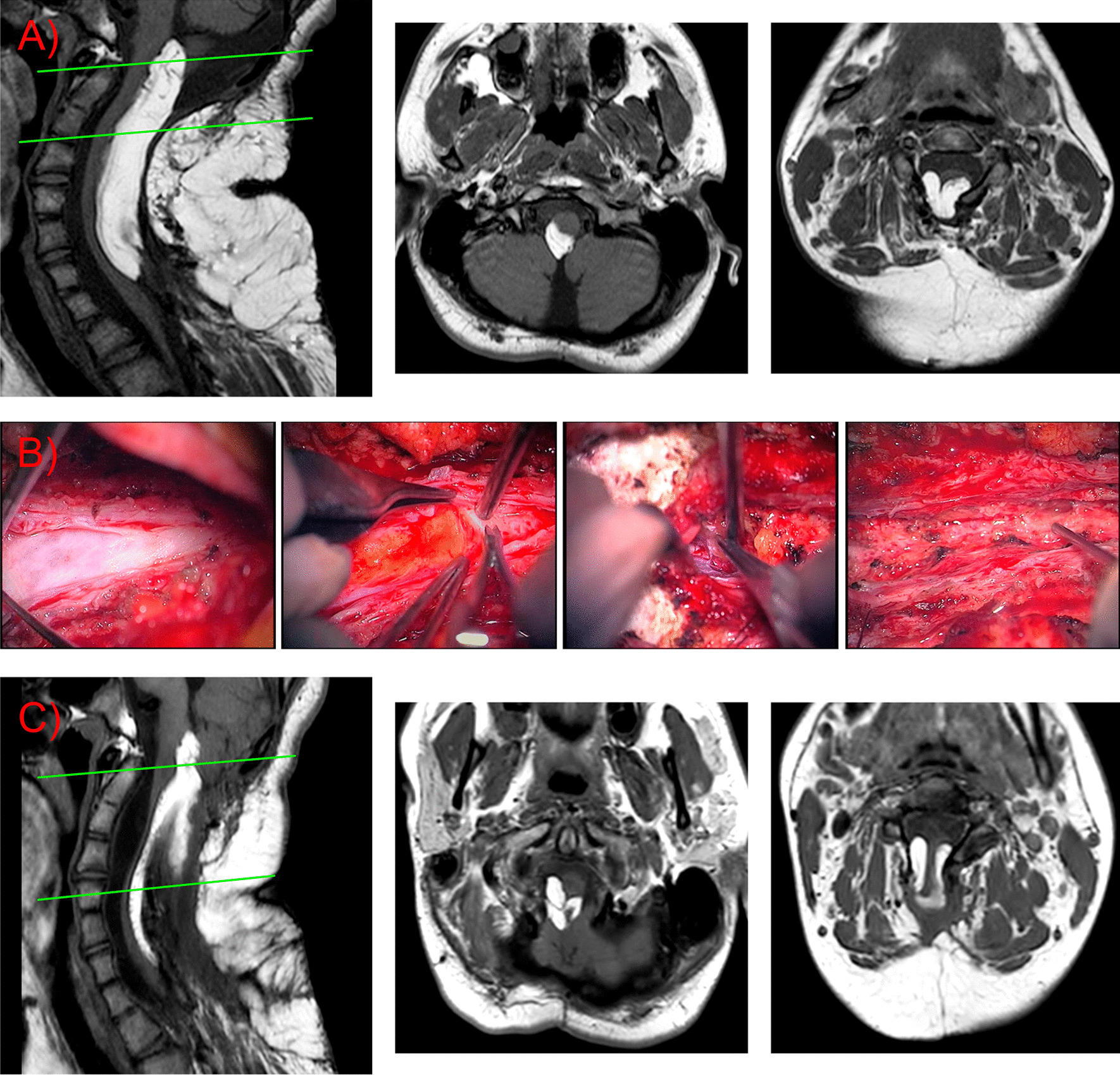
Case reports: Two patients, a 19-year-old South American woman and a 14-year-old boy with spinal lipomas, underwent surgical resection. The lipomas were not associated with dysraphism and were located in the cervicothoracic and craniocervical junctions. In both cases, we decided to operate due to clinical progression; the former had a progressive natural course, and the latter experienced clinical worsening after recurrence from previous surgeries. The surgery took place with the assistance of neurophysiological monitoring and intraoperative ultrasound; a partial resection and medullary decompression were done, following the more recent recommendations.

Discussion: The natural history of these lesions is currently unknown due to their rarity and the heterogeneity in the long-term follow-up of previously reported cases. Although previous reports describe good outcomes after surgical resection, long follow-ups, especially in young subjects, may show differences in these outcomes with progression and recurrence. We contribute to this last piece of evidence by describing two more cases of progression and recurrence.

Lessons: Long-term close follow-up should be done in young subjects with spinal lipomas, as they are more prone to an aggressive course. Metabolism and hormonal changes may be behind this progression. Reoperation must be considered if neurological decline is detected.

Keywords: Dysraphism; Lipomas; Malformation; Metabolism; Recurrence; Spine.


Conflict of interest statement

The authors declare that they have no competing interests.

Figures

Fig. 1
Fig. 1
Subjects chronological timelines
Fig. 2
Fig. 2
A Magnetic resonance images showing a spinal lipoma that went from the cervical region (C5) to the thoracic region (T5). Note the axial T2-weighted image (T2WI). This sequence confirmed the intramedullary subpial localization of the tumor. B Intraoperative images taken under the surgical microscope. Note the lipoma localization under a subpial coat, and the tumor remains anchored to the posterior part of the spinal cord. C Postoperative magnetic resonance imaging immediately after the surgical procedure and 2 years after surgery. No radiological progression is shown during this period, with the tumor remaining invariable. Green lines show the level where the sagittal magnetic resonance imaging is sliced to show the axial view
Fig. 3
Fig. 3
A Sagittal and axial Magnetic resonance images showing the cervicomedullary lipoma. Note the absence of laminas in the cervical vertebrae, a result of the previous surgical resections. B Intraoperative images. From left to right, we can see the arachnoid coat, the subpial lipoma, the cervicomedullary junction after lifting part of the lipoma, and finally, stimulation of residual tumor remains anchored to the posterior part of the medulla. C Postoperative MR images. The posterior lengthening of the medullary cord made resection of the tumor quite challenging. Green lines show the level where the sagittal magnetic resonance imaging is sliced to show the axial view

References

    1. Chen KY, Osorio J, Rivera J, Chou D. Intramedullary and extramedullary thoracic spinal lipomas without spinal dysraphism: clinical presentation and surgical management. World Neurosurg. 2019;121:156–159. doi: 10.1016/j.wneu.2018.09.156. - DOI - PubMed
    1. Şanh AM, Türkoǧlu E, Kahveci R, Šekerci Z. Intradural lipoma of the cervicothoracic spinal cord with intracranial extension. Child’s Nerv Syst. 2010;26(6):847–852. doi: 10.1007/s00381-009-1077-2. - DOI - PubMed
    1. Fleming KL, Davidson L, Gonzalez-Gomez I, Mccomb JG. Nondysraphic pediatric intramedullary spinal cord lipomas: report of 5 cases. J Neurosurg Pediatr. 2010;5(2):172–178. doi: 10.3171/2009.9.PEDS09238. - DOI - PubMed
    1. Bhatoe HS, Singh P, Chaturvedi A, Sahai K, Dutta V, Sahoo PK. Nondysraphic intramedullary spinal cord lipomas: a review. Neurosurg Focus. 2005;18(2):1–5. doi: 10.3171/foc.2005.18.2.10. - DOI - PubMed
    1. Lee M, Rezai AR, Abbott R, Coelho DH, Epstein FJ. Intramedullary spinal cord lipomas. J Neurosurg. 1995;82(3):394–400. doi: 10.3171/jns.1995.82.3.0394. - DOI - PubMed

Publication types

LinkOut - more resources