. 2023 Jul 24;14(1):180.
doi: 10.1186/s13287-023-03408-2.

Dendritic cells mediated by small extracellular vesicles derived from MSCs attenuated the ILC2 activity via PGE2 in patients with allergic rhinitis

Affiliations

Dendritic cells mediated by small extracellular vesicles derived from MSCs attenuated the ILC2 activity via PGE2 in patients with allergic rhinitis

Xiao-Qing Liu et al. Stem Cell Res Ther. .

Abstract

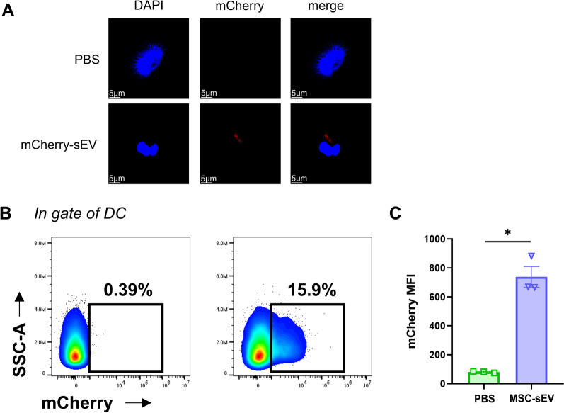
Background: Mesenchymal stromal cells-derived small extracellular vesicles (MSC-sEVs) have recently attracted considerable attention because of their therapeutic potential in various immune diseases. We previously reported that MSC-sEVs could exert immunomodulatory roles in allergic airway inflammation by regulating group 2 innate lymphoid cell (ILC2) and dendritic cell (DC) functions. Therefore, this study aimed to investigate the indirect effects of MSC-sEVs on ILC2s from patients with allergic rhinitis (AR) via DCs.

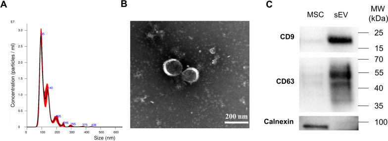
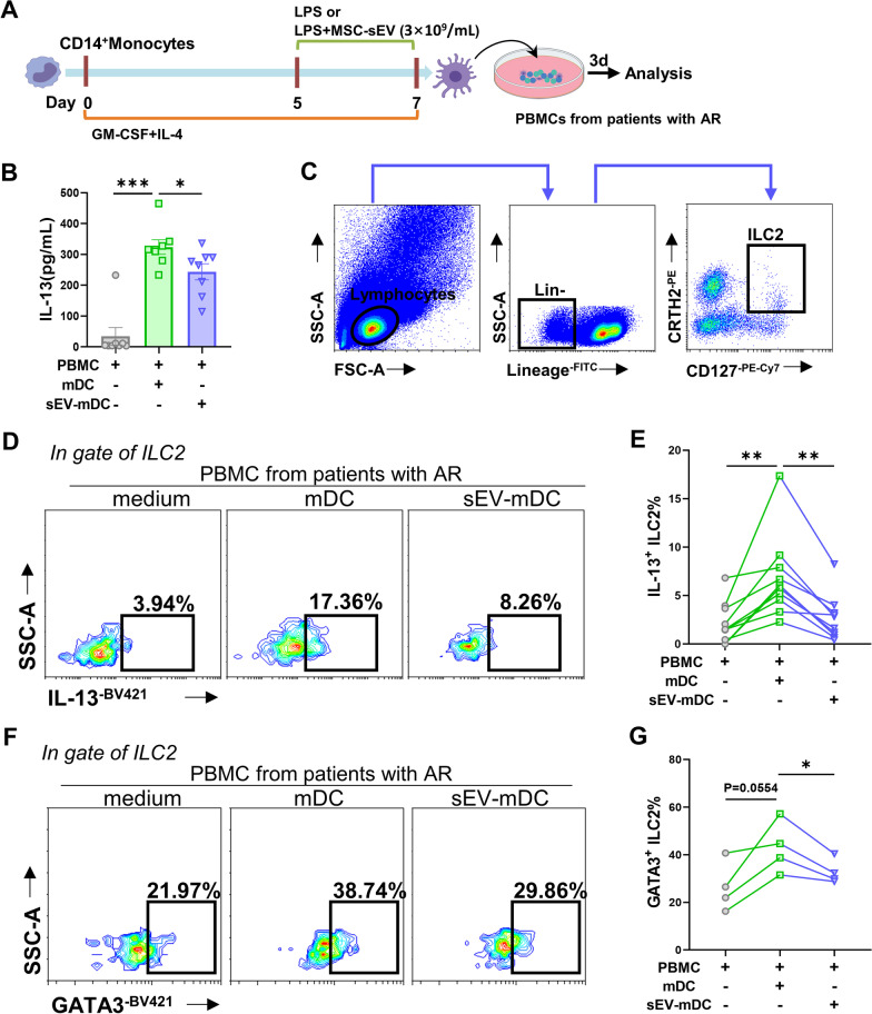
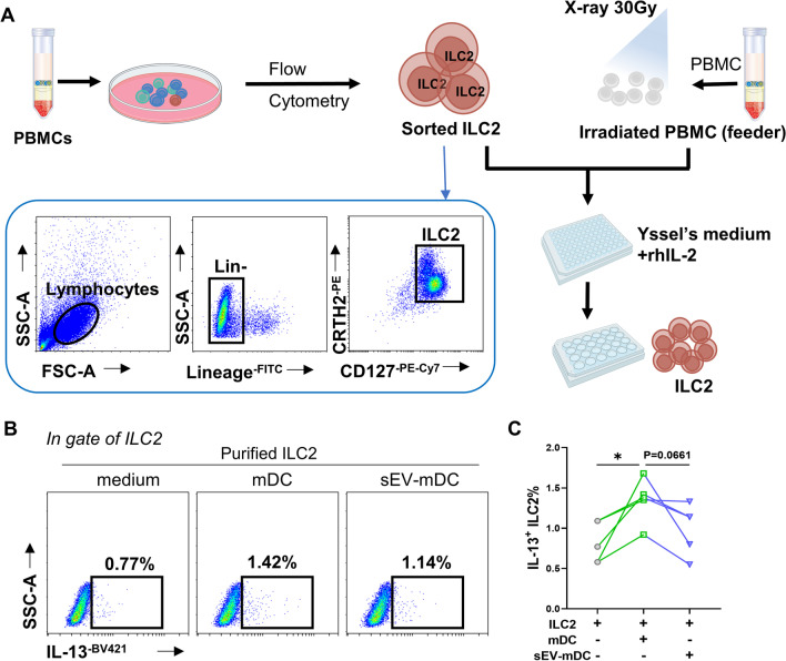
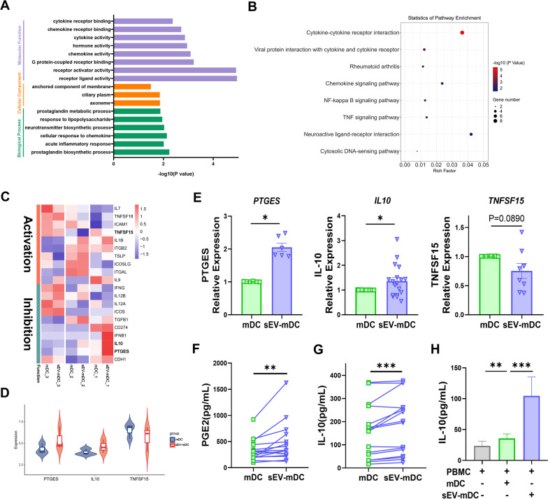
Methods: Here, we isolated sEVs from induced pluripotent stem cells-MSCs using anion-exchange chromatography and mature DCs (mDCs) were treated with MSC-sEVs. sEV-mDCs were co-cultured with peripheral blood mononuclear cells from patients with AR or purified ILC2s. The levels of IL-13 and GATA3 in ILC2s were examined by flow cytometry. Bulk RNA sequence for mDCs and sEV-mDCs was employed to further probe the potential mechanisms, which were then validated in the co-culture systems.

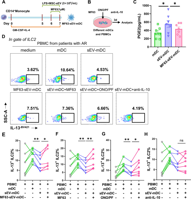
Results: sEV-mDCs showed impaired capacity in priming the levels of IL-13 and GATA3 in ILC2s when compared with mDCs. Furthermore, there was higher PGE2 and IL-10 production from sEV-mDCs, and the blockade of them especially the former one reversed the inhibitory effects of sEV-mDCs.

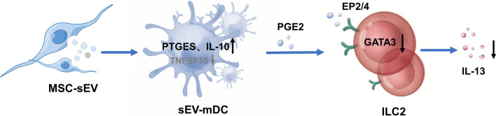
Conclusions: We demonstrated that MSC-sEVs were able to dampen the activating effects of mDCs on ILC2s in patients with AR. Mechanismly, the PGE2-EP2/4 axis played an essential role in the immunomodulatory effects of sEV-mDCs on ILC2s. Herein, we provided new insights into the mechanism underlying the therapeutic effects of MSC-sEVs in allergic airway inflammation.

Keywords: Allergic rhinitis; Dendritic cells; Group 2 innate lymphoid cells; Mesenchymal stromal cells; Prostaglandin E2; Small extracellular vesicles.


Conflict of interest statement

The authors declare no conflicts of interest in this work.

Figures

Fig. 1
Fig. 1
Identification of MSC-sEV. MSC-sEVs were isolated from iPSC-MSCs using anion-exchange chromatography. A. Nanoparticle Tracking Analysis (NTA) showed the particle size distribution and concentration of MSC-sEVs. B. TEM showed morphology and size of MSC-sEVs. C. CD9, CD63 and calnexin expression in MSCs and sEVs were determined by western blotting. The original blots are presented in Additional file 1: Fig. S4. MSC: mesenchymal stromal cells; MW: molecular weight; sEV: small extracellular vesicles; TEM: transmission electron microscope. Scale bar, 200 nm
Fig. 2
Fig. 2
MSC-sEVs inhibited the effects of dendritic cells on ILC2s in patients with allergic rhinitis. A. Schematic representation of mDCs and sEV-mDCs generation and co-cultures. BF. PBMCs from patients with AR were co-cultured with allogeneic mDCs or sEV-mDCs for 3 days. B. The levels of IL-13 in the supernatants were analyzed by ELISA (n = 8). C. Gating strategy of human ILC2s with LinCRTH2+CD127+. D-E. Intracellular IL-13 levels in ILC2s were analyzed by flow cytometry (n = 10). F-G. Levels of GATA3 in ILC2s were analyzed by flow cytometry (n = 4). DC: dendritic cell; ILC2: group 2 innate lymphoid cell; MSC: mesenchymal stromal cell; sEV: small extracellular vesicle. Data are shown as mean ± SEM. *P < 0.05, **P < 0.01, ***P < 0.001
Fig. 3
Fig. 3
sEV-mDCs exhibited impaired capacity on purified ILC2 function. A. Schematic of isolation and amplifying purified ILC2s. BC. Purified ILC2s were co-cultured with allogeneic mDCs or sEV-mDCs for 3 days. Intracellular IL-13 levels in ILC2s were analyzed by flow cytometry (n = 5). *P < 0.05
Fig. 4
Fig. 4
Uptake of MSC-sEV by DCs. DCs on day 5 were co-cultured with mCherry-labeled MSC-sEVs for 12 h. A. The cells were photographed by confocal microscopy. Panels showed mCherry (Red) and DAPI (Blue). BC. mCherry in DCs was analyzed using flow cytometry. MFI: mean fluorescence intensity. Scale bar, 5 μm. Data are shown as mean ± SEM. *P < 0.05
Fig. 5
Fig. 5
The expression of PGE2 and IL-10 in sEV-mDCs and mDCs. AB. GO analysis and KEGG enrichment analysis of differential mRNAs between mDCs and sEV-mDCs. C. Heatmap representation of differential mRNAs involved in ILC2s regulation between mDCs and sEV-mDCs. D. The violin plot showed the differential expression of PTGES, IL10 and TNFSF15 in mDCs and sEV-mDCs. E. The levels of PTGES, IL10 and TNFSF15 were examined by RT-quantitative PCR (n = 6–18). F. The levels of PGE2 in the supernatants of mDCs and sEV-mDCs (n = 16). G-H. The levels of IL-10 in the supernatants of mDCs and sEV-mDCs and co-cultured PBMCs (n = 13–16). PTGES: Prostaglandin E Synthase; IL-10: Interleukin-10; PGE2: Prostaglandin E2. Data are shown as mean ± SEM. *P < 0.05, **P < 0.01, ***P < 0.001
Fig. 6
Fig. 6
PGE2 mediated the impaired function of sEV-mDCs on ILC2s through EP2/4. PBMCs from patients with AR were co-cultured with allogeneic mDCs, sEV-mDCs, MF63-pretreated sEV-mDCs or combined with MF63 (0.1 μM), ONO/PF (1 μM), anti-IL-10 (2 μg/mL), respectively, for 3 days. A–B. Schematic of MF63-sEV-mDCs induction and blocking experiments. C. The levels of PGE2 in the supernatants of mDCs, sEV-mDCs and MF63-sEV-mDCs (n = 7). D. Intracellular IL-13 levels in ILC2s under different condition were analyzed by flow cytometry. E–H. Percentage of IL-13+ILC2s in the culture conditions as described (n = 7). MF63: pharmacological inhibitor of prostaglandin E Synthase; ONO/PF: ONO-AE3-208 and PF-04418948, antagonists for EP2/EP4. Data are shown as mean ± SEM. *P < 0.05, **P < 0.01
Fig. 7
Fig. 7
Schema for MSC-sEVs indirectly suppress ILC2 function in allergic rhinitis through increasing production of PGE2 from sEV-mDCs

References

    1. Brozek JL, Bousquet J, Agache I, Agarwal A, Bachert C, Bosnic-Anticevich S, et al. Allergic rhinitis and its impact on asthma (ARIA) guidelines-2016 revision. J Allergy Clin Immun. 2017;140(4):950–958. doi: 10.1016/j.jaci.2017.03.050. - DOI - PubMed
    1. Licona-Limon P, Kim LK, Palm NW, Flavell RA. TH2, allergy and group 2 innate lymphoid cells. Nat Immunol. 2013;14(6):536–542. doi: 10.1038/ni.2617. - DOI - PubMed
    1. Samolinski B. Pathogenesis of allergic rhinitis. Pneumonol Alergol Pol. 2002;70(Suppl 1):49–52. - PubMed
    1. Halim TY, Steer CA, Matha L, Gold MJ, Martinez-Gonzalez I, McNagny KM, et al. Group 2 innate lymphoid cells are critical for the initiation of adaptive T helper 2 cell-mediated allergic lung inflammation. Immunity. 2014;40(3):425–435. doi: 10.1016/j.immuni.2014.01.011. - DOI - PMC - PubMed
    1. Drake LY, Iijima K, Kita H. Group 2 innate lymphoid cells and CD4+ T cells cooperate to mediate type 2 immune response in mice. Allergy. 2014;69(10):1300–1307. doi: 10.1111/all.12446. - DOI - PMC - PubMed

Publication types