

Isoliquiritigenin induces HMOX1 and GPX4-mediated ferroptosis in gallbladder cancer cells
- PMID: 37488674
- PMCID: PMC10508381
- DOI: 10.1097/CM9.0000000000002675
Isoliquiritigenin induces HMOX1 and GPX4-mediated ferroptosis in gallbladder cancer cells
Abstract
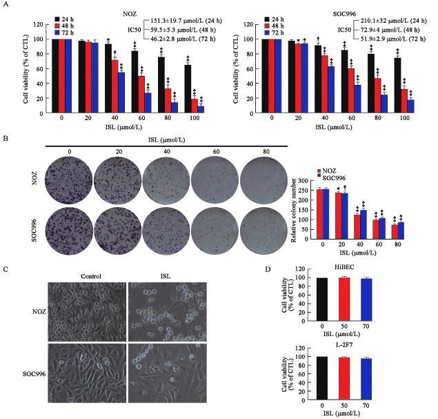
Background: Gallbladder cancer (GBC) is the most common malignant tumor of biliary tract. Isoliquiritigenin (ISL) is a natural compound with chalcone structure extracted from the roots of licorice and other plants. Relevant studies have shown that ISL has a strong anti-tumor ability in various types of tumors. However, the research of ISL against GBC has not been reported, which needs to be further investigated.
Methods: The effects of ISL against GBC cells in vitro and in vivo were characterized by cytotoxicity test, RNA-sequencing, quantitative real-time polymerase chain reaction, reactive oxygen species (ROS) detection, lipid peroxidation detection, ferrous ion detection, glutathione disulphide/glutathione (GSSG/GSH) detection, lentivirus transfection, nude mice tumorigenesis experiment and immunohistochemistry.
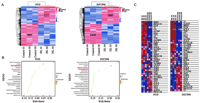
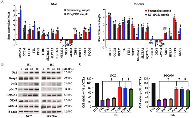
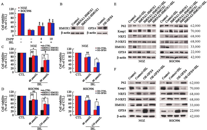
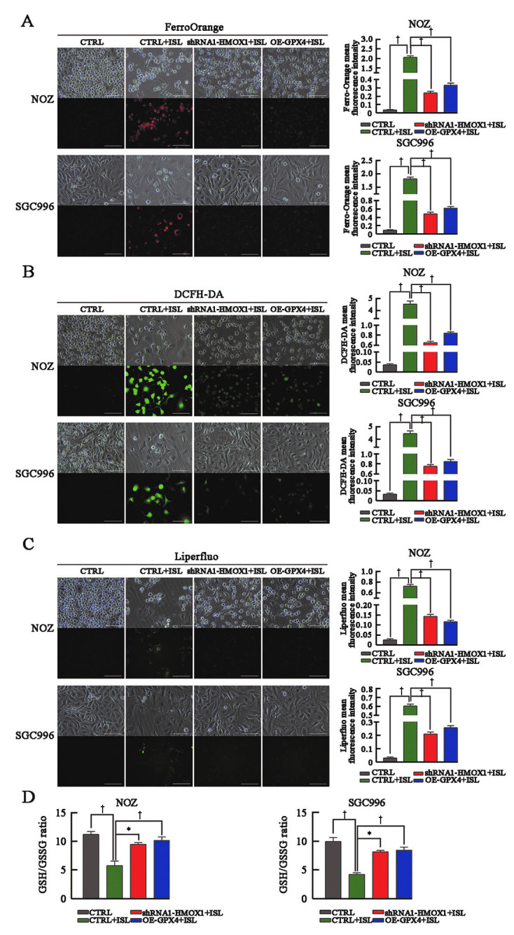
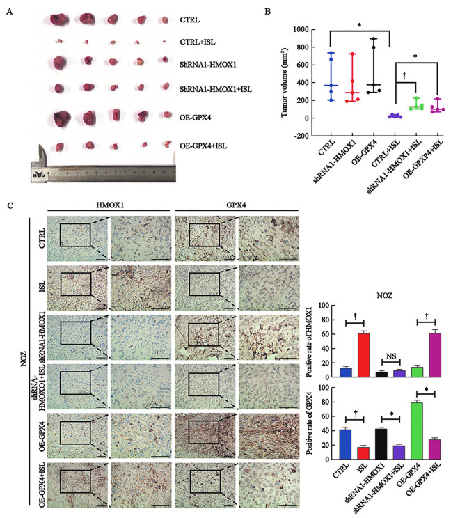
Results: ISL significantly inhibited the proliferation of GBC cells in vitro . The results of transcriptome sequencing and bioinformatics analysis showed that ferroptosis was the main pathway of ISL inhibiting the proliferation of GBC, and HMOX1 and GPX4 were the key molecules of ISL-induced ferroptosis. Knockdown of HMOX1 or overexpression of GPX4 can reduce the sensitivity of GBC cells to ISL-induced ferroptosis and significantly restore the viability of GBC cells. Moreover, ISL significantly reversed the iron content, ROS level, lipid peroxidation level and GSSG/GSH ratio of GBC cells. Finally, ISL significantly inhibited the growth of GBC in vivo and regulated the ferroptosis of GBC by mediating HMOX1 and GPX4 .
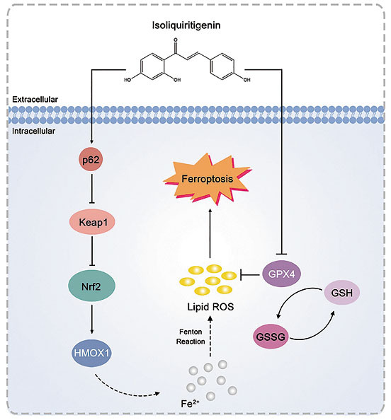
Conclusion: ISL induced ferroptosis in GBC mainly by activating p62-Keap1-Nrf2-HMOX1 signaling pathway and down-regulating GPX4 in vitro and in vivo . This evidence may provide a new direction for the treatment of GBC.
Copyright © 2023 The Chinese Medical Association, produced by Wolters Kluwer, Inc. under the CC-BY-NC-ND license.
Conflict of interest statement
None.
Figures
Similar articles
-
Isoliquiritigenin alleviates myocardial ischemia-reperfusion injury by regulating the Nrf2/HO-1/SLC7a11/GPX4 axis in mice.Free Radic Biol Med. 2024 Aug 20;221:1-12. doi: 10.1016/j.freeradbiomed.2024.05.012. Epub 2024 May 9. Free Radic Biol Med. 2024. PMID: 38734270
-
Dihydrotanshinone I inhibits gallbladder cancer growth by targeting the Keap1-Nrf2 signaling pathway and Nrf2 phosphorylation.Phytomedicine. 2024 Jul;129:155661. doi: 10.1016/j.phymed.2024.155661. Epub 2024 Apr 21. Phytomedicine. 2024. PMID: 38677269
-
Curcumol Enhances the Sensitivity of Gastric Cancer to Cisplatin Resistance by Inducing Ferroptosis Through the P62/KEAP1/NRF2 Pathway.Integr Cancer Ther. 2024 Jan-Dec;23:15347354241294043. doi: 10.1177/15347354241294043. Integr Cancer Ther. 2024. PMID: 39511708 Free PMC article.
-
[Kelch-like ech-associated protein 1/nuclear factor E2-related factor 2/antioxidant response element pathway alleviates ferroptosis in sepsis by regulating oxidative stress].Zhonghua Wei Zhong Bing Ji Jiu Yi Xue. 2021 Jul;33(7):881-884. doi: 10.3760/cma.j.cn121430-20210130-00180. Zhonghua Wei Zhong Bing Ji Jiu Yi Xue. 2021. PMID: 34412763 Review. Chinese.
-
Astragaloside IV regulates the ferroptosis signaling pathway via the Nrf2/SLC7A11/GPX4 axis to inhibit PM2.5-mediated lung injury in mice.Int Immunopharmacol. 2022 Nov;112:109186. doi: 10.1016/j.intimp.2022.109186. Epub 2022 Sep 15. Int Immunopharmacol. 2022. PMID: 36115280 Review.
Cited by
-
Theranostic Applications of Taurine-Derived Carbon Dots in Colorectal Cancer: Ferroptosis Induction and Multifaceted Antitumor Mechanisms.Int J Nanomedicine. 2025 Jun 16;20:7613-7635. doi: 10.2147/IJN.S516926. eCollection 2025. Int J Nanomedicine. 2025. PMID: 40546797 Free PMC article.
-
Impact of SPP1 and HMOX1 Genes in Glioma: Correlations With Oncolytic Virus Infection, Adverse Prognosis and Increased Cell Proliferation.J Cell Mol Med. 2025 Jun;29(11):e70651. doi: 10.1111/jcmm.70651. J Cell Mol Med. 2025. PMID: 40495644 Free PMC article.
-
Phytochemicals against Osteoarthritis by Inhibiting Apoptosis.Molecules. 2024 Mar 27;29(7):1487. doi: 10.3390/molecules29071487. Molecules. 2024. PMID: 38611766 Free PMC article. Review.
-
Programmed Cell Death-Related Gene Signature Associated with Prognosis and Immune Infiltration and the Roles of HMOX1 in the Proliferation and Apoptosis were Investigated in Uveal Melanoma.Genes Genomics. 2024 Jul;46(7):785-801. doi: 10.1007/s13258-024-01521-x. Epub 2024 May 20. Genes Genomics. 2024. PMID: 38767825 Free PMC article.
-
ITRAQ-based proteomics analysis of human ectopic endometrial stromal cells treated by Maqian essential oil.BMC Complement Med Ther. 2023 Nov 27;23(1):427. doi: 10.1186/s12906-023-04246-8. BMC Complement Med Ther. 2023. PMID: 38012607 Free PMC article.
References
-
- Li M Zhang Z Li X Ye J Wu X Tan Z, et al. . Whole-exome and targeted gene sequencing of gallbladder carcinoma identifies recurrent mutations in the ErbB pathway. Nat Genet 2014;46: 872–876. doi: 10.1038/ng.3030. - PubMed
-
- Zhang Y Zuo C Liu L Hu Y Yang B Qiu S, et al. . Single-cell RNA-sequencing atlas reveals an MDK-dependent immunosuppressive environment in ErbB pathway-mutated gallbladder cancer. J Hepatol 2021;75: 1128–1141. doi: 10.1016/j.jhep.2021.06.023. - PubMed
-
- Li Y, Li M, Liu Y. Current status and future prospects of basic research on gallbladder carcinoma. Chin J Pract Surgery 2021;41: 52–55. doi: 10.19538/j.cjps.issn1005-2208.2021.01.08
MeSH terms
Substances
LinkOut - more resources
Full Text Sources
Medical
