. 2023 Oct 1;34(10):1672-1686.
doi: 10.1681/ASN.0000000000000197. Epub 2023 Jul 25.

Elucidating the Proximal Tubule HNF4A Gene Regulatory Network in Human Kidney Organoids

Affiliations

Elucidating the Proximal Tubule HNF4A Gene Regulatory Network in Human Kidney Organoids

Yasuhiro Yoshimura et al. J Am Soc Nephrol. .

Abstract

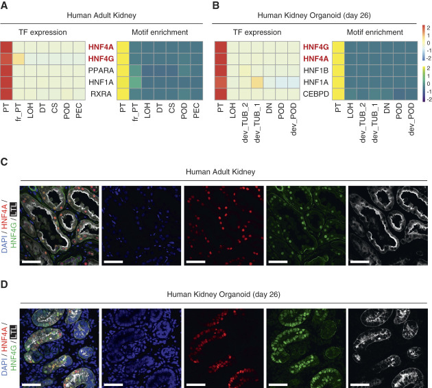
Significance statement: HNF4 genes promote proximal tubule differentiation in mice, but their function in human nephrogenesis is not fully defined. This study uses human pluripotent stem cell (PSC)-derived kidney organoids as a model to investigate HNF4A and HNF4G functions. The loss of HNF4A , but not HNF4G , impaired reabsorption-related molecule expression and microvilli formation in human proximal tubules. Cleavage under targets and release using nuclease (CUT&RUN) sequencing and CRISPR-mediated transcriptional activation (CRISPRa) further confirm that HNF4A directly regulates its target genes. Human kidney organoids provide a good model for studying transcriptional regulation in human kidney development.

Background: The proximal tubule plays a major role in electrolyte homeostasis. Previous studies have shown that HNF4A regulates reabsorption-related genes and promotes proximal tubule differentiation during murine kidney development. However, the functions and gene regulatory mechanisms of HNF4 family genes in human nephrogenesis have not yet been investigated.

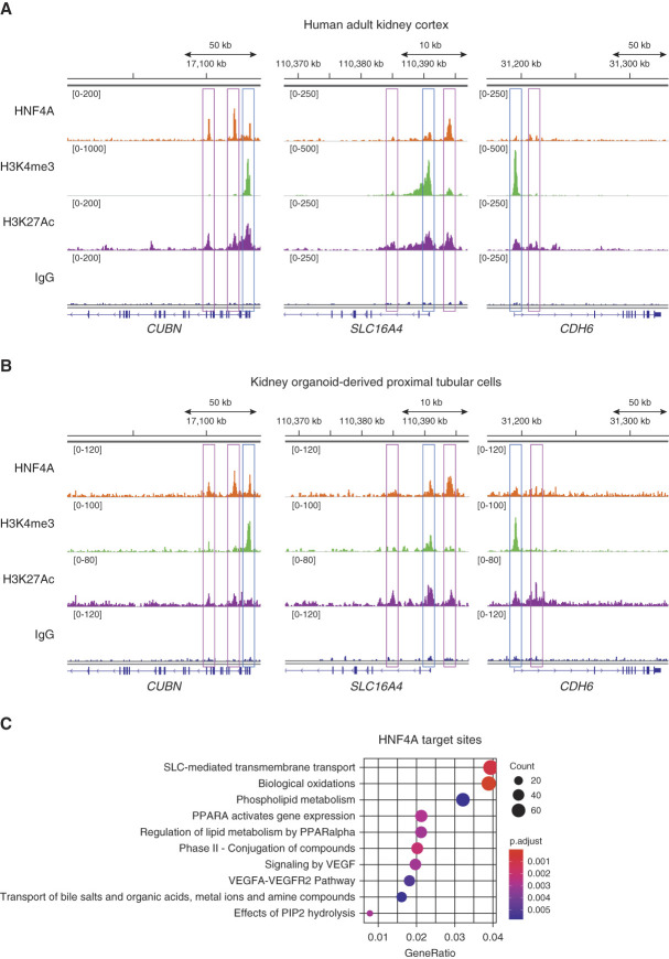
Methods: We generated HNF4A -knock out (KO), HNF4G -KO, and HNF4A/4G -double KO human pluripotent stem cell lines, differentiated each into kidney organoids, and used immunofluorescence analysis, electron microscopy, and RNA-seq to analyze them. We probed HNF4A-binding sites genome-wide by cleavage under targets and release using nuclease sequencing in both human adult kidneys and kidney organoid-derived proximal tubular cells. Clustered Regularly Interspaced Short Palindromic Repeats-mediated transcriptional activation validated HNF4A and HNF4G function in proximal tubules during kidney organoid differentiation.

Results: Organoids lacking HNF4A , but not HNF4G , showed reduced expression of transport-related, endocytosis-related, and brush border-related genes, as well as disorganized brush border structure in the apical lumen of the organoid proximal tubule. Cleavage under targets and release using nuclease revealed that HNF4A primarily bound promoters and enhancers of genes that were downregulated in HNF4A -KO, suggesting direct regulation. Induced expression of HNF4A or HNF4G by CRISPR-mediated transcriptional activation drove increased expression of selected target genes during kidney organoid differentiation.

Conclusions: This study reveals regulatory mechanisms of HNF4A and HNF4G during human proximal tubule differentiation. The experimental strategy can be applied more broadly to investigate transcriptional regulation in human kidney development.


Conflict of interest statement

B.D. Humphreys reports Consultancy: Chinook Therapeutics, Janssen, Pfizer; Ownership Interest: Chinook Therapeutics; Research Funding: Janssen, Pfizer; Honoraria: Novartis; Patents or Royalties: AG, Evotec; and Advisory or Leadership Role: Seminars in Nephrology: Editorial Board, Kidney International: Editorial Board, JCI Insight: Editorial Board, American Journal of Physiology Renal Physiology, Editorial Board, RegMed XB, Regenerative Medicine Crossing Borders: SAB, ASCI: President, Chinook Therapeutics, SAB, Member of Board of Scientific Advisors of NIDDK. J.H. Miner reports Consultancy: Bayer, Guidepoint; Research Funding: Chinook Therapeutics, Keros Therapeutics, Lung Therapeutics Inc, Myonid Therapeutics; Honoraria: AstraZeneca, Axiom Healthcare Strategies, Pfizer, Visterra; Patents or Royalties: Elsevier; Kerafast; Maze Therapeutics; Advisory or Leadership Role: American Society for Matrix Biology, President; Journal of Clinical Investigation, Consulting Editor; Kidney International, Editorial Board; Matrix Biology, Editorial Board; Matrix Biology Plus, Editorial Board; and Other Interests or Relationships: Alport Syndrome Foundation (Scientific Advisory Research Network). All remaining authors have nothing to disclose.

Figures

None
Graphical abstract
Figure 1
Figure 1
HNF4A and HNF4G are top regulatory transcription factors in the human proximal tubular cells. (A, B) Heatmaps showing gene expression levels (left) and motif enrichment in ATAC peaks (right) of top listed transcription factors in the proximal tubular cells of multiome datasets generated from human adult kidneys (A) and kidney organoids (B). (C, D) Immunofluorescence images of HNF4A (red), HNF4G (green), LTL (white), and nuclear DAPI (blue) staining of human adult kidney (C) and day 26 kidney organoid (D). Scale bars indicate 50 µm. CS, connecting segment; dev_POD, developing podocyte; dev_TUB, developing tubule; DN, distal nephron; DT, distal tubule; fr_PT, failed repaired proximal tubule; LOH, Loop of Henle; PEC, parietal epithelial cells in the glomerulus; POD, podocyte; PT, proximal tubule.
Figure 2
Figure 2
Kidney organoid induction from HNF4A-KO, HNF4G-KO, and HNF4A/HNF4G-DKO iPSC lines. (A, B) Strategy for targeting human HNF4A (A) and HNF4G (B) genes with CRISPR gene editing to generate HNF4A-KO and HNF4G-KO iPSC lines, respectively. The sequencing at the targeted sites of the HNF4A-KO (A) and HNF4G-KO (B) iPSC lines are shown. Magenta letters and hyphens in the sequencing indicate inserted or deleted bases, respectively. (C) Immunofluorescence images of CDH1 (red, distal nephron epithelium marker), WT1 (green, podocyte marker), LTL (white, proximal tubular cell marker), and nuclear DAPI (blue) staining of day 26 human kidney organoids induced from wild-type (clone AN1.1), HNF4A-KO (clone 2F2), HNF4G-KO (clone 2D3), and HNF4A/4G-KO (clone 3) iPSC lines. Scale bars indicate 200 µm. (D) Immunofluorescence images of HNF4A (red), HNF4G (green), LTL (white), and nuclear DAPI (blue) staining of day 26 human kidney organoids induced from wild-type (clone AN1.1), HNF4A-KO (clone 2G4), HNF4G-KO (clone 2B3), and HNF4A/4G-KO (clone 5) iPSC lines. Scale bars indicate 50 µm.
Figure 3
Figure 3
HNF4A-KO and HNF4A/4G-DKO organoids showed downregulation of transport-related, endocytosis-related, and brush border–related genes. (A, C, E) Volcano plots showing the differentially expressed genes in the comparison of HNF4A-KO (clone 2G4, A), HNF4G-KO (clone 2D3, C), and HNF4A/4G-DKO (clone 5, E) kidney organoids to wild-type (clone AN1.1) kidney organoids. (B, D, F) Enriched pathways from gene ontology analysis of the downregulated genes in the HNF4A-KO (clone 2G4, B), HNF4G-KO (clone 2D3, D), and HNF4A/4G-DKO (clone 5, F) kidney organoids compared with wild-type (clone AN1.1) kidney organoids. Each clone shows an average of different batches of organoids (n=6). Brown bars, biological process (BP); yellow bars, cellular component (CC); green bars, molecular function (MF).
Figure 4
Figure 4
HNF4A-KO and HNF4A/4G-DKO organoids showed disorganized microvilli structure in the proximal tubule. (A) Immunofluorescence images of CUBN (red), HNF4A (green), LTL (white), and nuclear DAPI (blue) staining of day 26 human kidney organoids induced from wild-type (clone AN1.1), HNF4A-KO (clone 2G4), HNF4G-KO (clone 2D3), and HNF4A/4G-KO (clone 5) iPSC lines. Scale bars indicate 50 µm. (B) Transmission electron microscopic images of day 26 human kidney organoids induced from wild-type (clone AN1.1), HNF4A-KO (clone 2G4), HNF4G-KO (clone 2D3), and HNF4A/4G-KO (clone 5) iPSC lines. Lower panels are magnified images of the yellow boxes in their corresponding upper images. Blue arrowheads indicate disorganized microvilli. Scale bars indicate 10 µm (upper panels) and 500 nm (lower panels).
Figure 5
Figure 5
CUT&RUN sequencing confirmed direct regulation of HNF4A for transport and endocytosis-related genes in the human kidneys. (A, B) Integrative genomics viewer (IGV) images showing target sites of HNF4A (top), H3K4me3 (promoter activity, second from the top), H3K27Ac (enhancer and promoter activity, second from the bottom), and IgG (negative control, bottom) antibodies in CUT&RUN sequencing with human adult kidney cortex (A) and kidney organoid-derived proximal tubule (B) of CUBN (left), SLC16A4 (middle), and CDH6 (right) genes. Blue boxes indicate promoter regions; magenta boxes indicate enhancer regions. (C) Enriched pathways from Reactome pathway analysis of the HNF4A-binding peaks detected in the CUT&RUN analysis of human adult kidney cortex.
Figure 6
Figure 6
CRISPR activation using kidney organoid system confirmed transcriptional activity differences between HNF4A and HNF4G. (A) Schematic of experiment for CRISPR activation (CRISPRa) during kidney organoid differentiation. (B, C) qRT-PCR results of CRISPRa kidney organoids (day 26) targeting HNF4A (B) and HNF4G (C). n=3 biological replicates. The data are presented as mean±SEM. * P < 0.05, ** P < 0.01. (D) Immunofluorescence images of SLC3A1 (magenta), HNF4A (yellow), and nuclear DAPI (blue) staining of CRISPRa kidney organoids targeting HNF4A without DOX (left) and with DOX (right) conditions. Scale bars indicate 100 μm. hES cells, human embryonic stem cells; n.s., not significant.

References

    1. Takaori K, Nakamura J, Yamamoto S, Nakata H, Sato Y, Takase M. Severity and frequency of proximal tubule injury determines renal prognosis. J Am Soc Nephrol. 2016;27(8):2393–2406. doi:10.1681/ASN.2015060647 - DOI - PMC - PubMed
    1. Kirita Y, Wu H, Uchimura K, Wilson PC, Humphreys BD. Cell profiling of mouse acute kidney injury reveals conserved cellular responses to injury. Proc Natl Acad Sci. 2020;117(27):15874–15883. doi:10.1073/pnas.2005477117 - DOI - PMC - PubMed
    1. Gerhardt LMS, Liu J, Koppitch K, Cippà PE, McMahon AP. Single-nuclear transcriptomics reveals diversity of proximal tubule cell states in a dynamic response to acute kidney injury. Proc Natl Acad Sci USA. 2021;118(27):e2026684118. doi:10.1073/pnas.2026684118 - DOI - PMC - PubMed
    1. Kobayashi A, Valerius MT, Mugford JW, Carroll TJ, Self M, Oliver G. Six2 defines and regulates a multipotent self-renewing nephron progenitor population throughout mammalian kidney development. Cell Stem Cell. 2008;3(2):169–181. doi:10.1016/j.stem.2008.05.020 - DOI - PMC - PubMed
    1. Costantini F, Kopan R. Patterning a complex organ: branching morphogenesis and nephron segmentation in kidney development. Dev Cell. 2010;18(5):698–712. doi:10.1016/j.devcel.2010.04.008 - DOI - PMC - PubMed

Publication types

Substances