. 2024 Feb;19(2):447-457.
doi: 10.4103/1673-5374.373669.

BMPRII+ neural precursor cells isolated and characterized from organotypic neurospheres: an in vitro model of human fetal spinal cord development

Affiliations

BMPRII+ neural precursor cells isolated and characterized from organotypic neurospheres: an in vitro model of human fetal spinal cord development

Michael W Weible Ii et al. Neural Regen Res. 2024 Feb.

Abstract

Roof plate secretion of bone morphogenetic proteins (BMPs) directs the cellular fate of sensory neurons during spinal cord development, including the formation of the ascending sensory columns, though their biology is not well understood. Type-II BMP receptor (BMPRII), the cognate receptor, is expressed by neural precursor cells during embryogenesis; however, an in vitro method of enriching BMPRII+ human neural precursor cells (hNPCs) from the fetal spinal cord is absent. Immunofluorescence was undertaken on intact second-trimester human fetal spinal cord using antibodies to BMPRII and leukemia inhibitory factor (LIF). Regions of highest BMPRII+ immunofluorescence localized to sensory columns. Parenchymal and meningeal-associated BMPRII+ vascular cells were identified in both intact fetal spinal cord and cortex by co-positivity with vascular lineage markers, CD34/CD39. LIF immunostaining identified a population of somas concentrated in dorsal and ventral horn interneurons, mirroring the expression of LIF receptor/CD118. A combination of LIF supplementation and high-density culture maintained culture growth beyond 10 passages, while synergistically increasing the proportion of neurospheres with a stratified, cytoarchitecture. These neurospheres were characterized by BMPRII+/MAP2ab+/-/βIII-tubulin+/nestin-/vimentin-/GFAP-/NeuN- surface hNPCs surrounding a heterogeneous core of βIII-tubulin+/nestin+/vimentin+/GFAP+/MAP2ab-/NeuN- multipotent precursors. Dissociated cultures from tripotential neurospheres contained neuronal (βIII-tubulin+), astrocytic (GFAP+), and oligodendrocytic (O4+) lineage cells. Fluorescence-activated cell sorting-sorted BMPRII+ hNPCs were MAP2ab+/-/βIII-tubulin+/GFAP-/O4- in culture. This is the first isolation of BMPRII+ hNPCs identified and characterized in human fetal spinal cords. Our data show that LIF combines synergistically with high-density reaggregate cultures to support the organotypic reorganization of neurospheres, characterized by surface BMPRII+ hNPCs. Our study has provided a new methodology for an in vitro model capable of amplifying human fetal spinal cord cell numbers for > 10 passages. Investigations of the role BMPRII plays in spinal cord development have primarily relied upon mouse and rat models, with interpolations to human development being derived through inference. Because of significant species differences between murine biology and human, including anatomical dissimilarities in central nervous system (CNS) structure, the findings made in murine models cannot be presumed to apply to human spinal cord development. For these reasons, our human in vitro model offers a novel tool to better understand neurodevelopmental pathways, including BMP signaling, as well as spinal cord injury research and testing drug therapies.

Keywords: BMPRII; bone morphogenetic protein; histotypic; human spinal cord development; leukemia inhibitory factor; neurosphere; organotypic; reaggregate; sensory columns.


Conflict of interest statement

None

Figures

Figure 1
Figure 1
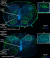
Human fetal spinal cord BMPRII immunostaining at gestational ages of 15–16 weeks. Cryostat section (12 μm) of the human fetal spinal cord estimated to be at gestational ages of 15–16 weeks showing intense BMPRII+ immunoreactivity (green) along the ventral surface, as well as the dorsal (sensory) and ventral (motor) horns. Insets A–G shown at higher magnification, scale bar: 50 μm in D; nuclei counterstain DAPI (blue). Staining was observed along the dorsal surface and the sensory and motor neurons in the dorsal and ventral horns. The dotted red line demarcates regions of higher intensity BMPRII-immunoreactivity in dorsal horns. Magenta marks the outer margin of higher-intensity BMPRII-immunoreactivity in the ventral funiculus, while orange demarcates the extent of the structure. (A) BMPRII+ immunoreactivity in the dorsal horn (B) BMPRII+ immunoreactivity in the ventral horn (C) BMPRII+ immunoreactivity at the ventral funiculus. (D–F) Vascular tube-like structures showed strong immunoreactivity to BMPRII staining, while (G) shows an example of staining of the meninges surrounding the spinal cord. The main anatomical structures are identified by labeling. 5× magnification, scale bar: 200 μm. BMP: Bone morphogenetic protein; BMPRII: type-II BMP receptor; DAPI: 4′,6-diamidino-2-phenylindole.
Figure 2
Figure 2
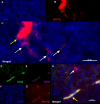
BMPRII+ hNPCs (red) can also be identified in the human fetal brain cortex (A–C) and are identified by a lack of vascular marker expression (D–H). Scattered BMPRII+ putative hNPCs (white arrows) are present in the parenchyma of a second-trimester human fetal brain cortex (B), nuclei counterstain DAPI (blue; A), shown merged in (C). By virtue of lower expression, these cells are clearly distinct from BMPRII+ cells in vascular tubes (yellow arrow). 63× magnification, scale bar: 20 μm. Quadruple-labeled immunofluorescence of an intact second-trimester spinal cord, utilizing antibodies against BMPRII (G) combined with two established vascular lineage markers (CD34; green; D) and ecto-ADPase CD39 (magenta; E), showing a colocalized, triple-labeled vascular segment (yellow arrow in H). BMPRII (red) is highly expressed by vascular cells (yellow arrow), while scattered BMPRII+ single cells (putative hNPCs) negative for vascular markers are evident in the tissue parenchyma (e.g., white arrow in (H); merge of all images). Nuclear counterstain DAPI (blue; F) labels nuclei. 63× magnification, scale bar: 20 μm. BMP: Bone morphogenetic protein; BMPRII: type-II BMP receptor; DAPI: 4′,6-diamidino-2-phenylindole; hNPCs: human neural precursor cells.
Figure 3
Figure 3
Human fetal spinal cord LIFR and LIF immunostaining at gestational ages of 14–15 weeks. Upper section: Cryostat section (12 μm) of human fetal spinal cord estimated to be at gestational ages of 14–15 weeks showing LIF immunoreactivity (green) mostly within ventral horn motor interneurons (dotted white line and inset, A; scale bar in inset 100 μm). Some positive staining was also observed in the dorsal horn sensory interneurons (dotted red outline). The dorsal columns, lateral and ventral funiculus were negative. Immunopositivity was also seen in the peripheral edge of the section. Lower section: LIFR staining (green) was similar to LIF with scattered positive immunoreactivity within ventral horn motor interneurons (labeled). Positive staining was also observed in the dorsal horn sensory interneurons (dotted red outline and inset, B; scale bar in inset 100 μm). The dorsal columns, lateral and ventral funiculus were negative. Nuclear counterstain DAPI (blue). The main anatomical structures are identified by labeling: 5× magnification, scale bar for both sections: 200 μm. DAPI: 4′,6-Diamidino-2-phenylindole; LIF: leukemia inhibitory factor; LIFR: LIF receptor.
Figure 4
Figure 4
LIF positively regulates neurosphere growth as a function of passage number. (A) First-generation floating neural aggregates often have refractive bubbles on their surfaces. Scale bar: 50 μm. (B) Neural aggregates expanded at low density in media supplemented with LIF (empty shapes) undergo an increase in cell proliferation versus untreated cultures (filled shapes). LIF: Leukemia inhibitory factor.
Figure 5
Figure 5
Effect of culture conditions and LIF prolong culture expansion over repeated passages, while maintaining tripotentiality. (A) High-density reaggregation in the presence of LIF permits the prolonged expansion of cultures compared with the low-density passage. (B) Aggregates (P0) and reaggregates (P1) were fixed, sectioned, and subjected to immunofluorescence staining with antibodies to nestin (green), glial fibrillary acidic protein (GFAP, red), βIII-tubulin (blue), and nuclear counterstain DAPI (magenta). Emergent small neural aggregates were homogeneous, while reaggregates were recomposed of numerous neural phenotypes. (C) Free-floating reaggregates were allowed to expand in size and grossly resemble normal aggregate spheres, including the possible presence of microspikes along the surface. Reaggregates similar to that represented in C were dissociated, plated, fixed, and subjected to immunofluorescence staining with antibodies to GFAP (red), βIII-tubulin (green), O4 (blue), and DAPI (magenta). (D) The proportion of each phenotype was quantified. Data are expressed as the mean ± SEM. (E–H) Heterogeneous reaggregate neurospheres give rise to multiple neural phenotypes. Images in F–H are shown merged and at higher magnification in E. (E) βIII-tubulin+/GFAP NPC (βIII-tubulin+/GFAP: broad diffuse nuclei, epithelial-like morphology (up arrow) or compact nuclei, bipolar/neuronal-morphology (down arrows), GFAP+ NPC (βIII-tubulin+/GFAP+, broad diffuse nuclei, epithelial-like morphology) and OPC (O4+/GFAP compact dense nuclei, multipolar morphology). DAPI: 4′,6-Diamidino-2-phenylindole; LIF: leukemia inhibitory factor; NPC: neural precursor cell; OPC: oligodendrocyte precursor cell.
Figure 6
Figure 6
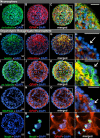
Immunocharacterization of organotypic reaggregate neurospheres. (A–D) Neurospheres were identified as having a heterogeneous composition, using antibodies against βIII-tubulin (green) and GFAP (red). The images in A and B are shown merged in C and box magnified in D. The neurosphere shows no obvious organization of neural cells. White boxes in C, G, K, and O indicate areas shown zoomed in D, H, L, and P, respectively (scale bars: 25 μm). (E–P) A surface layer of βIII-tubulin-expressing hNPCs on organotypic reaggregate neurospheres. (E–H) The surface cells (arrow) do not express the neural stem cell markers nestin (green) or vimentin (red). The images in (E–F) are shown merged in G, box magnified in H, where a surface layer of nestin/vimentin cells is clearly evident. (I–L) Labeling with βIII-tubulin (green) and negative for GFAP (red) in the outer layer of cells demonstrated the stratification of organotypic reaggregate neurospheres. The images in I and J are shown merged in K and box magnified in L. The classical βIII-tubulin perinuclear staining (arrow) found on the surface cells is often indicative of lineage elaboration towards an immature neuronal phenotype, GFAP expression is also absent from these surface cells. (M–P) Most surface cells of organotypic reaggregate neurospheres have not begun to express the mature neuronal marker MAP2ab (green, arrow in P). The images in M–N are shown merged in O and the box is magnified in P. (Q) All reaggregates were negative for NeuN staining (green), a protein up-regulated in postsynaptic neurons. (R–T) However, reaggregates plated on extracellular matrix-coated glass (as per methods) can generate cells that mature into NeuN-expressing neurons (R). (S) GFAP staining (red) of the same area as R. (T) Merge of R and S. All nuclei were counterstained with DAPI (blue), and scale bars are as indicated. DAPI: 4′,6-Diamidino-2-phenylindole; GFAP: glial fibrillary acidic protein; hNPC: human neural precursor cell; MAP: microtubule-associated protein; NeuN: neuron-specific nuclear protein.
Figure 7
Figure 7
Effects of tissue culture condition on organotypic reaggregate neurosphere phenotype. (A) A comparison of sphere size showed that uniform stratification significantly favored populations of medium size spheres (200–500 μm diameter, e.g., J, Figure 5C) compared with small (< 200 μm diameter, e.g. F, Figure 5B) and large spheres (> 500 μm diameter). ***P < 0.001. (B) The effects of high-density reaggregation compared with low-density aggregation in combination with the presence or absence of LIF were assayed for their effects on neurosphere stratification. Both high-density and LIF-containing culture conditions were significantly different from low-density spheres or those grown in the absence of LIF. (C–F) Immunocharacterization of organotypic reaggregate neurospheres. Labeling with βIII-tubulin (C, green) shows an outer layer of immunoreactivity typical of organotypic neurospheres, while being negative for GFAP (D, red). The inside of organotypic neurospheres shows heterogenous βIII-tubulin and GFAP labeling. Co-staining with BMPRII (E) confirmed that βIII-tubulin+ cells are double positive; merge shown in (F), and white boxed areas in C–F are zoomed below each sphere to show the fine detail of expressing cells. Reaggregate neurospheres were also characterized by positive immunoreactivity for LIF (G) and LIFR (H) but with no obvious indication of stratification; whereas endogenous BMP (I) and BMPRII (J) (all green) were both localized to the cell surface layer. All nuclei were counterstained with DAPI (magenta or blue, as indicated). Scale bar in F and J is 25 μm, and in F (inset below), 12.5 μm. BMP: Bone morphogenetic protein; BMPRII: type-II BMP receptor; DAPI: 4′,6-diamidino-2-phenylindole; GFAP: glial fibrillary acidic protein; LIF: leukemia inhibitory factor; LIFR: LIF receptor; ORN: organotypic reaggregate neurosphere.
Figure 8
Figure 8
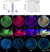
Characterization of fluorescence-assisted cell sorting-sorted BMPRII+ hNPCs. (A–E) Sorted cells were plated, fixed, and subjected to immunofluorescence staining with antibodies to O4 (blue), GFAP (red), MAP2ab (green), and the nuclei counterstained with DAPI (magenta). Image (A) shows sorted cells at low magnification. Images in C through E are shown with polarized bright field and merged at a higher magnification in B (from the area of the white box in A). (F) A representative single-label FACS histogram plot of fluorescence intensity versus cell number for collected BMPRII+ cells (green trace) and control (2° antibody only; red trace). “Collected” shows the fluorescence intensity gate used to collect the BMPRII+ cells. (G) Sorted cells were plated and the proportion of each phenotype was quantified. A significant increase was found for the number of MAP2ab+ cells from approximately 24% in control to 83% in collected. (H) Cells were characterized using antibodies against each indicated marker where ‘+’ indicates > 90% of the cells with neuronal morphology were positive and ‘–’ indicates < 10% of these cells were positive. Data are expressed as the mean ± SEM, and scale bars are as indicated. BMP: Bone morphogenetic protein; BMPRII: type-II BMP receptor; DAPI: 4′,6-diamidino-2-phenylindole; GFAP: glial fibrillary acidic protein; hNPCs: human neural precursor cells; MAP: microtubule-associated protein; NeuN: neuron-specific nuclear protein; PSA-NCAM: polysialylated-neural cell adhesion molecule.

Similar articles

Cited by

References

    1. Andrews MG, Del Castillo LM, Ochoa-Bolton E, Yamauchi K, Smogorzewski J, Butler SJ. BMPs direct sensory interneuron identity in the developing spinal cord using signal-specific not morphogenic activities. Elife. (2017);6:e30647. - PMC - PubMed
    1. Andrews MG, Kong J, Novitch BG, Butler SJ. New perspectives on the mechanisms establishing the dorsal-ventral axis of the spinal cord. Curr Top Dev Biol. (2019);132:417–450. - PMC - PubMed
    1. Andruska A, Spiekerkoetter E. Consequences of BMPR2 deficiency in the pulmonary vasculature and beyond:contributions to pulmonary arterial hypertension. Int J Mol Sci. (2018);19:2499. - PMC - PubMed
    1. Atkinson C, Stewart S, Upton PD, Machado R, Thomson JR, Trembath RC, Morrell NW. Primary pulmonary hypertension is associated with reduced pulmonary vascular expression of type II bone morphogenetic protein receptor. Circulation. (2002);105:1672–1678. - PubMed
    1. Back SA, Luo NL, Borenstein NS, Volpe JJ, Kinney HC. Arrested oligodendrocyte lineage progression during human cerebral white matter development:dissociation between the timing of progenitor differentiation and myelinogenesis. J Neuropathol Exp Neurol. (2002);61:197–211. - PubMed