

Suppression of MUC1-Overexpressing Tumors by a Novel MUC1/CD3 Bispecific Antibody
- PMID: 37489369
- PMCID: PMC10366937
- DOI: 10.3390/antib12030047
Suppression of MUC1-Overexpressing Tumors by a Novel MUC1/CD3 Bispecific Antibody
Abstract
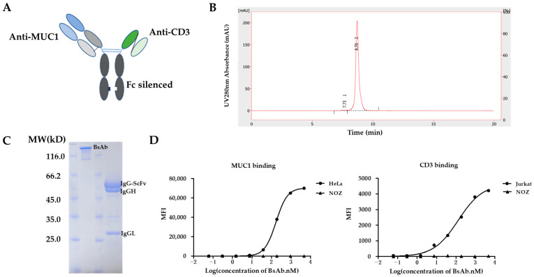
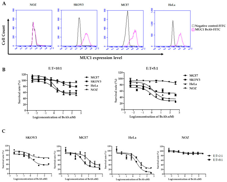
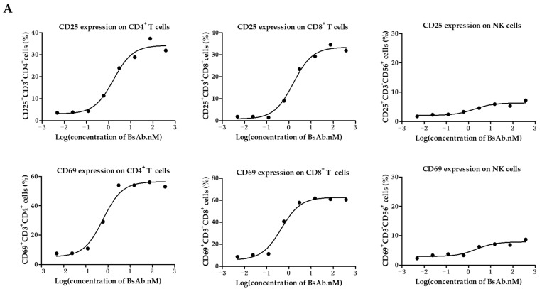
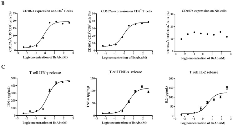
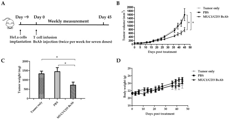
Mucin1 (MUC1) is abnormally glycosylated and overexpressed in a variety of epithelial cancers and plays a critical role in tumor progression. MUC1 has received remark attention as an oncogenic molecule and is considered a valuable tumor target for immunotherapy, while many monoclonal antibodies (mAbs) targeting MUC1-positive cancers in clinical studies lack satisfactory results. It would be highly desirable to develop an effective therapy against MUC1-expressing cancers. In this study, we constructed a novel T cell-engaging bispecific antibody (BsAb) targeting MUC1 and CD3 with the Fab-ScFv-IgG format. A high quality of MUC1-CD3 BsAb can be acquired through a standard method. Our study suggested that this BsAb could specifically bind to MUC1- and CD3-positive cells and efficiently enhance T cell activation, cytokine release, and cytotoxicity. Furthermore, our study demonstrated that this BsAb could potently redirect T cells to eliminate MUC1-expressing tumor cells in vitro and significantly suppress MUC1-positive tumor growth in a xenograft mouse model. Thus, T cell-engaging MUC1/CD3 BsAb could be an effective therapeutic approach to combat MUC1-positive tumors and our MUC1/CD3 BsAb could be a promising candidate in clinical applications for the treatment of MUC1-positive cancer patients.
Keywords: BsAb; Mucin1; epithelial cancers; immunotherapy; oncogenic molecule.
Conflict of interest statement
S.L. and H.Y. are employees of BenHealth Biopharmaceutical (Shenzhen) Co., Ltd. The study was supported by BenHealth Biopharmaceutical (Shenzhen) Co., Ltd. The funder was not involved in the study design, collection, analysis, interpretation of data, writing of this article, or decision to submit it for publication. The company and this cooperation did not affect the authenticity and objectivity of the experimental results of this work.
Figures
Similar articles
-
MUC1-specific targeting immunotherapy with bispecific antibodies: inhibition of xenografted human bile duct carcinoma growth.Cancer Res. 1996 Sep 15;56(18):4205-12. Cancer Res. 1996. PMID: 8797593
-
A Novel Bispecific Antibody Targeting CD3 and Lewis Y with Potent Therapeutic Efficacy against Gastric Cancer.Biomedicines. 2021 Aug 20;9(8):1059. doi: 10.3390/biomedicines9081059. Biomedicines. 2021. PMID: 34440263 Free PMC article.
-
A T-cell-engaging B7-H4/CD3-bispecific Fab-scFv Antibody Targets Human Breast Cancer.Clin Cancer Res. 2019 May 1;25(9):2925-2934. doi: 10.1158/1078-0432.CCR-17-3123. Epub 2019 Feb 8. Clin Cancer Res. 2019. PMID: 30737243
-
Recent advances of bispecific antibodies in solid tumors.J Hematol Oncol. 2017 Sep 20;10(1):155. doi: 10.1186/s13045-017-0522-z. J Hematol Oncol. 2017. PMID: 28931402 Free PMC article. Review.
-
Performance of CD3xCD19 bispecific monoclonal antibodies in B cell malignancy.Leuk Lymphoma. 1995 Nov;19(5-6):381-93. doi: 10.3109/10428199509112195. Leuk Lymphoma. 1995. PMID: 8590837 Review.
Cited by
-
Targeting Siglec-Sialylated MUC1 Immune Axis in Cancer.Cancers (Basel). 2024 Mar 29;16(7):1334. doi: 10.3390/cancers16071334. Cancers (Basel). 2024. PMID: 38611013 Free PMC article. Review.
-
The Role of MUC1 in Renal Cell Carcinoma.Biomolecules. 2024 Mar 7;14(3):315. doi: 10.3390/biom14030315. Biomolecules. 2024. PMID: 38540735 Free PMC article. Review.
-
Mechanisms of Response and Tolerance to Active RAS Inhibition in KRAS-Mutant Non-Small Cell Lung Cancer.Cancer Discov. 2024 Nov 1;14(11):2183-2208. doi: 10.1158/2159-8290.CD-24-0421. Cancer Discov. 2024. PMID: 38975897 Free PMC article.
-
MUC1 and MUC16: critical for immune modulation in cancer therapeutics.Front Immunol. 2024 Feb 1;15:1356913. doi: 10.3389/fimmu.2024.1356913. eCollection 2024. Front Immunol. 2024. PMID: 38361923 Free PMC article. Review.
-
Development of CAR-T Therapies and Personalized Vaccines for the Treatment of Cholangiocarcinoma: Current Progress, Mechanisms of Action, and Challenges.Am J Pathol. 2025 Mar;195(3):453-469. doi: 10.1016/j.ajpath.2024.10.021. Epub 2024 Dec 14. Am J Pathol. 2025. PMID: 39675505 Review.
References
LinkOut - more resources
Full Text Sources
Research Materials
Miscellaneous
