. 2023 Aug 29;148(9):765-777.
doi: 10.1161/CIRCULATIONAHA.122.062646. Epub 2023 Jul 25.

Detection of Left Ventricular Systolic Dysfunction From Electrocardiographic Images

Affiliations

Detection of Left Ventricular Systolic Dysfunction From Electrocardiographic Images

Veer Sangha et al. Circulation. .

Abstract

Background: Left ventricular (LV) systolic dysfunction is associated with a >8-fold increased risk of heart failure and a 2-fold risk of premature death. The use of ECG signals in screening for LV systolic dysfunction is limited by their availability to clinicians. We developed a novel deep learning-based approach that can use ECG images for the screening of LV systolic dysfunction.

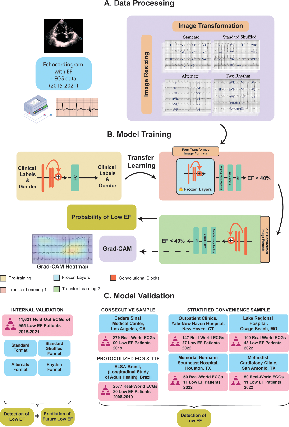
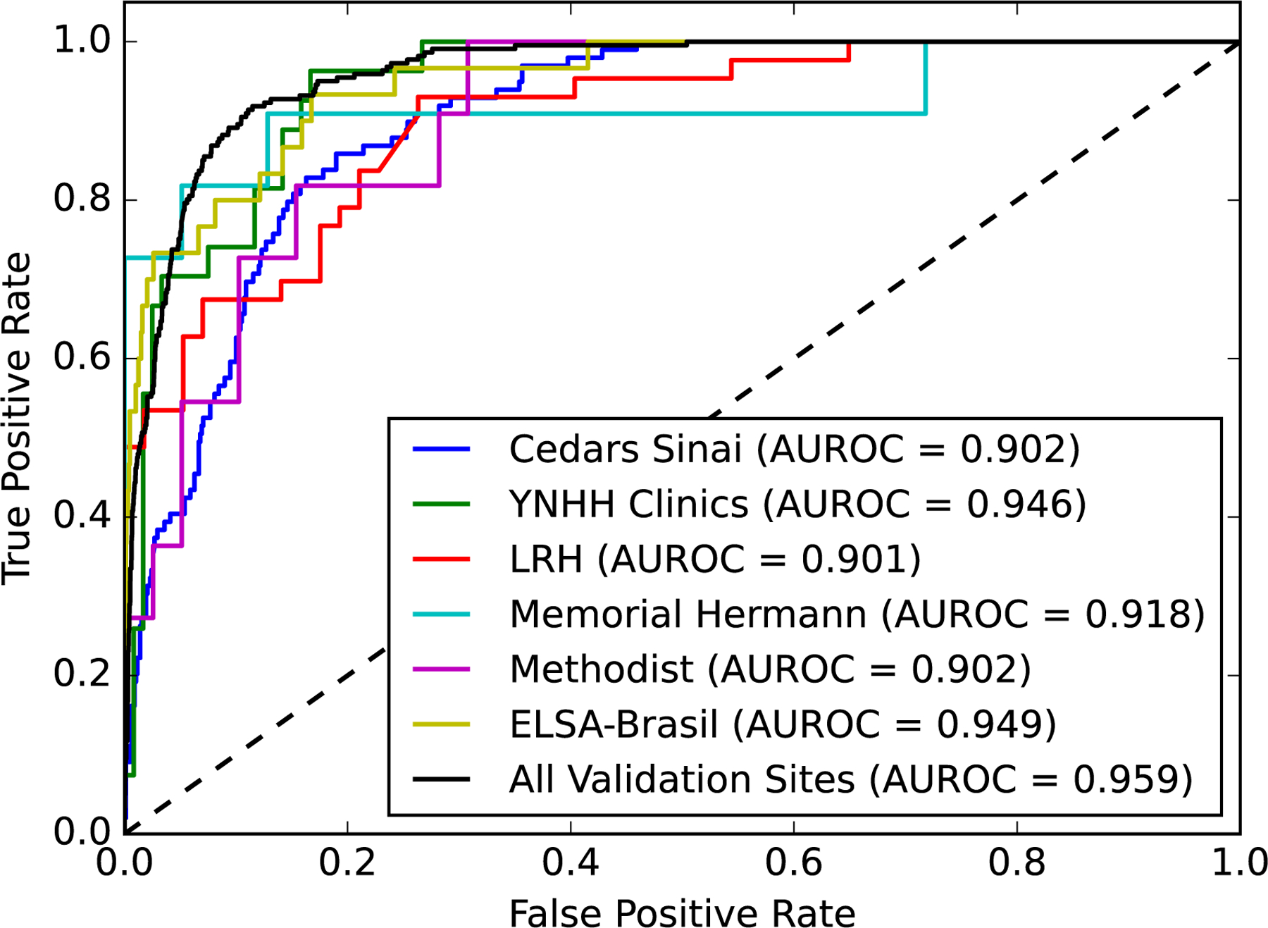
Methods: Using 12-lead ECGs plotted in multiple different formats, and corresponding echocardiographic data recorded within 15 days from the Yale New Haven Hospital between 2015 and 2021, we developed a convolutional neural network algorithm to detect an LV ejection fraction <40%. The model was validated within clinical settings at Yale New Haven Hospital and externally on ECG images from Cedars Sinai Medical Center in Los Angeles, CA; Lake Regional Hospital in Osage Beach, MO; Memorial Hermann Southeast Hospital in Houston, TX; and Methodist Cardiology Clinic of San Antonio, TX. In addition, it was validated in the prospective Brazilian Longitudinal Study of Adult Health. Gradient-weighted class activation mapping was used to localize class-discriminating signals on ECG images.

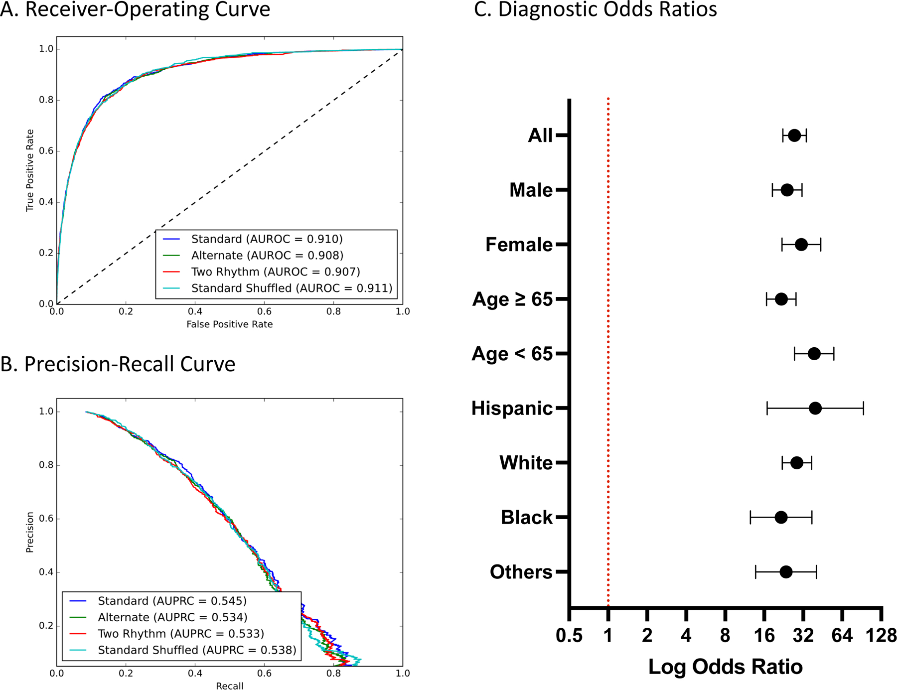
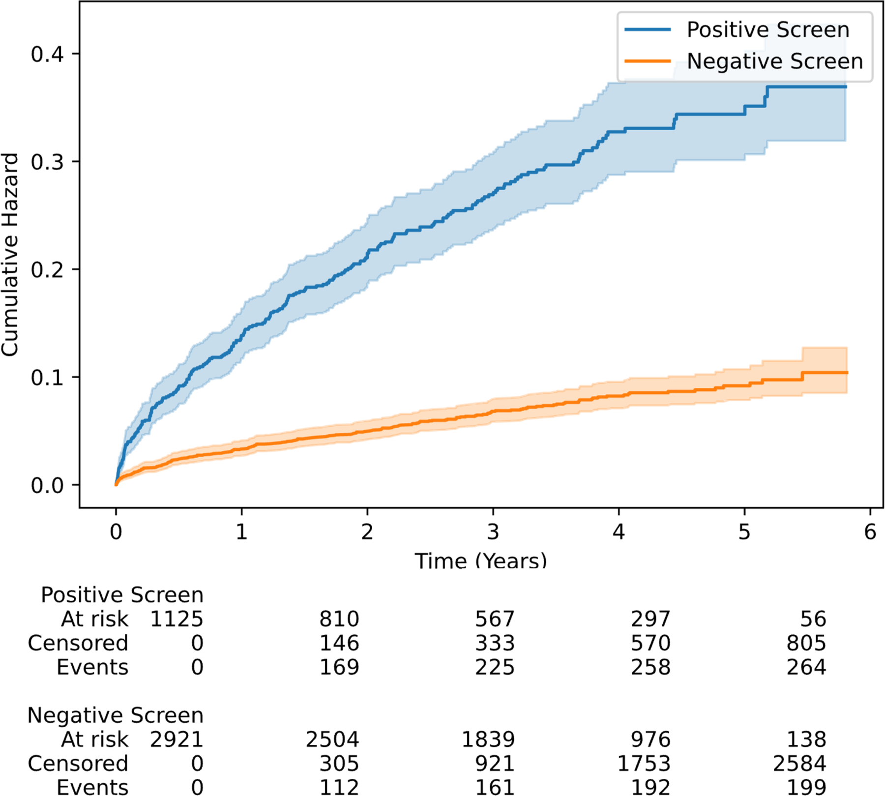
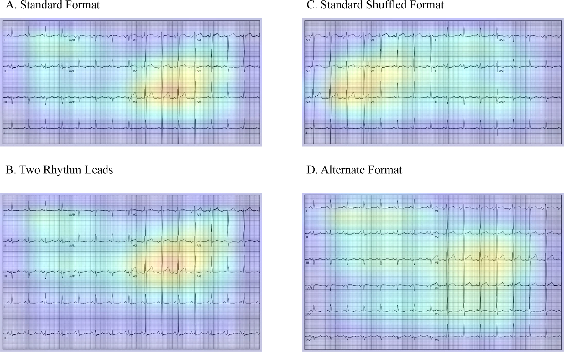
Results: Overall, 385 601 ECGs with paired echocardiograms were used for model development. The model demonstrated high discrimination across various ECG image formats and calibrations in internal validation (area under receiving operation characteristics [AUROCs], 0.91; area under precision-recall curve [AUPRC], 0.55); and external sets of ECG images from Cedars Sinai (AUROC, 0.90 and AUPRC, 0.53), outpatient Yale New Haven Hospital clinics (AUROC, 0.94 and AUPRC, 0.77), Lake Regional Hospital (AUROC, 0.90 and AUPRC, 0.88), Memorial Hermann Southeast Hospital (AUROC, 0.91 and AUPRC 0.88), Methodist Cardiology Clinic (AUROC, 0.90 and AUPRC, 0.74), and Brazilian Longitudinal Study of Adult Health cohort (AUROC, 0.95 and AUPRC, 0.45). An ECG suggestive of LV systolic dysfunction portended >27-fold higher odds of LV systolic dysfunction on transthoracic echocardiogram (odds ratio, 27.5 [95% CI, 22.3-33.9] in the held-out set). Class-discriminative patterns localized to the anterior and anteroseptal leads (V2 and V3), corresponding to the left ventricle regardless of the ECG layout. A positive ECG screen in individuals with an LV ejection fraction ≥40% at the time of initial assessment was associated with a 3.9-fold increased risk of developing incident LV systolic dysfunction in the future (hazard ratio, 3.9 [95% CI, 3.3-4.7]; median follow-up, 3.2 years).

Conclusions: We developed and externally validated a deep learning model that identifies LV systolic dysfunction from ECG images. This approach represents an automated and accessible screening strategy for LV systolic dysfunction, particularly in low-resource settings.

Keywords: artificial intelligence; biomedical technology; electrocardiography; heart failure; machine learning; ventricular dysfunction, left.


Conflict of interest statement

Disclosures Dr Mortazavi reported receiving grants from the National Institute of Biomedical Imaging and Bioengineering, National Heart, Lung, and Blood Institute, US Food and Drug Administration, and the US Department of Defense Advanced Research Projects Agency outside the submitted work; in addition, Dr Mortazavi has a pending patent on predictive models using electronic health records (US20180315507A1). Dr A.H. Ribeiro is funded by the Kjell och Märta Beijer Foundation. Dr Krumholz works under contract with the Centers for Medicare & Medicaid Services to support quality measurement programs, was a recipient of a research grant from Johnson & Johnson, through Yale University, to support clinical trial data sharing; was a recipient of a research agreement, through Yale University, from the Shenzhen Center for Health Information for work to advance intelligent disease prevention and health promotion; collaborates with the National Center for Cardiovascular Diseases in Beijing; receives payment from the Arnold & Porter Law Firm for work related to the Sanofi clopidogrel litigation, from the Martin Baughman Law Firm for work related to the Cook Celect IVC filter litigation, and from the Siegfried and Jensen Law Firm for work related to Vioxx litigation; chairs a cardiac scientific advisory board for UnitedHealth; was a member of the IBM Watson Health Life Sciences board; is a member of the advisory board for element science, the advisory board for Facebook, and the physician advisory board for Aetna; and is co-founder of Hugo Health, a personal health information platform, and co-founder of Refactor Health, a health care artificial intelligence–augmented data management company. Dr A.L.P. Ribeiro is supported in part by CNPq (465518/2014-1, 310790/2021-2, and 409604/2022-4) and by FAPEMIG (PPM-00428-17, RED-00081-16, and PPE-00030-21). V. Sangha and Dr Khera are the coinventors of US provisional patent application No. 63/346,610, “Articles and methods for format-independent detection of hidden cardiovascular disease from printed electrocardiographic images using deep learning.” Dr Khera receives support from the National Heart, Lung, and Blood Institute of the National Institutes of Health (under award K23HL153775) and the Doris Duke Charitable Foundation (under award 2022060). He receives support from the Blavatnik Foundation through the Blavatnik fund for innovation at Yale. He also receives research support, through Yale, from Bristol-Myers Squibb, and Novo Nordisk. He is an associate editor for JAMA. In addition to 63/346,610, Dr Khera is a coinventor of US provisional patent applications 63/177,117, 63/428,569, and 63/484,426. He is also a founder of Evidence2Health, a precision health platform to improve evidence-based cardiovascular care.

Figures

Figure 1.
Figure 1.. Study Outline A) Data processing, B) Model training, and C) Model validation.
Abbreviations: ECG, electrocardiogram; EF, ejection fraction; FC, fully connected layers; Grad-CAM, gradient-weighted class activation mapping; CT, Connecticut; ELSA-Brasil, Estudo Longitudinal de Saúde do Adulto (The Brazilian Longitudinal Study of Adult Health); MO, Missouri; TX, Texas. *The transfer learning strategy in developing the current model includes transferring model initialization weights from the previous algorithm originally trained to detect cardiac rhythm disorders and the hidden label of gender from ECG images. The transfer learning was used as initialization weights for the EfficientNet B3 convolutional neural network being trained to detect LV systolic dysfunction. Other than the weights, clinical and gender labels were not input into the current model.
Figure 2.
Figure 2.. Model Performance Measures A) Receiver-Operating and B) Precision-Recall Curves on images in held-out test set C) Diagnostic Odds Ratios across age, gender, and race subgroups on standard format images in the held-out test set.
Abbreviations: AUROC, area under receiver-operating characteristic curve; AUPRC, area under precision-recall curve.
Figure 3.
Figure 3.
Cumulative hazard curves for incident LV systolic dysfunction in model-predicted positive and negative screens amongst the members of the held-out test set with LVEF ≥ 40% and at least one follow-up measurement.
Figure 4.
Figure 4.. Gradient-weighted Class Activation Mapping (Grad-CAMs) across ECG formats. A) Standard format B) Two rhythm leads C) Standard shuffled format D) Alternate format.
The heatmaps represent averages of the 100 positive cases with the most confident model predictions for LVEF < 40%.
Figure 5.
Figure 5.. Receiver-Operating Curves for external validation sites.
Abbreviations: AUROC, area under receiver-operating characteristic curve; EF, Ejection fraction; LRH, Lake Regional Hospital; YNHH, Yale New Haven Hospital

References

    1. Wang TJ, Evans JC, Benjamin EJ, Levy D, LeRoy EC, Vasan RS. Natural history of asymptomatic left ventricular systolic dysfunction in the community. Circulation. 2003;108:977–982. - PubMed
    1. Srivastava PK, DeVore AD, Hellkamp AS, Thomas L, Albert NM, Butler J, Patterson JH, Spertus JA, Williams FB, Duffy CI, Hernandez AF, Fonarow GC. Heart Failure Hospitalization and Guideline-Directed Prescribing Patterns Among Heart Failure With Reduced Ejection Fraction Patients. JACC Heart Fail. 2021;9:28–38. - PubMed
    1. Wolfe NK, Mitchell JD, Brown DL. The independent reduction in mortality associated with guideline-directed medical therapy in patients with coronary artery disease and heart failure with reduced ejection fraction. Eur Heart J Qual Care Clin Outcomes. 2021;7:416–421. - PMC - PubMed
    1. Heidenreich PA, Bozkurt B, Aguilar D, Allen LA, Byun JJ, Colvin MM, Deswal A, Drazner MH, Dunlay SM, Evers LR, Fang JC, Fedson SE, Fonarow GC, Hayek SS, Hernandez AF, Khazanie P, Kittleson MM, Lee CS, Link MS, Milano CA, Nnacheta LC, Sandhu AT, Stevenson LW, Vardeny O, Vest AR, Yancy CW. 2022 AHA/ACC/HFSA Guideline for the Management of Heart Failure: A Report of the American College of Cardiology/American Heart Association Joint Committee on Clinical Practice Guidelines. Circulation. 2022;101161CIR0000000000001063. - PubMed
    1. Wang TJ, Levy D, Benjamin EJ, Vasan RS. The epidemiology of “asymptomatic” left ventricular systolic dysfunction: implications for screening. Ann Intern Med 2003;138:907–916. - PubMed

Publication types