. 2023 Aug;37(8):e23103.
doi: 10.1096/fj.202300862R.

Chromatin architectural factor CTCF is essential for progesterone-dependent uterine maturation

Affiliations

Chromatin architectural factor CTCF is essential for progesterone-dependent uterine maturation

Sylvia C Hewitt et al. FASEB J. 2023 Aug.

Abstract

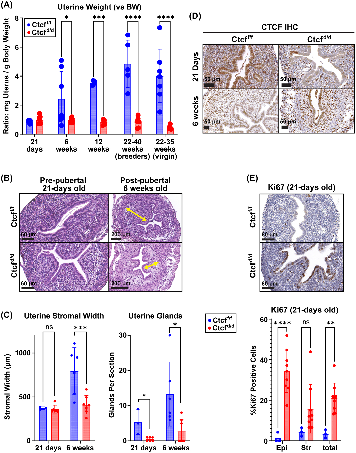
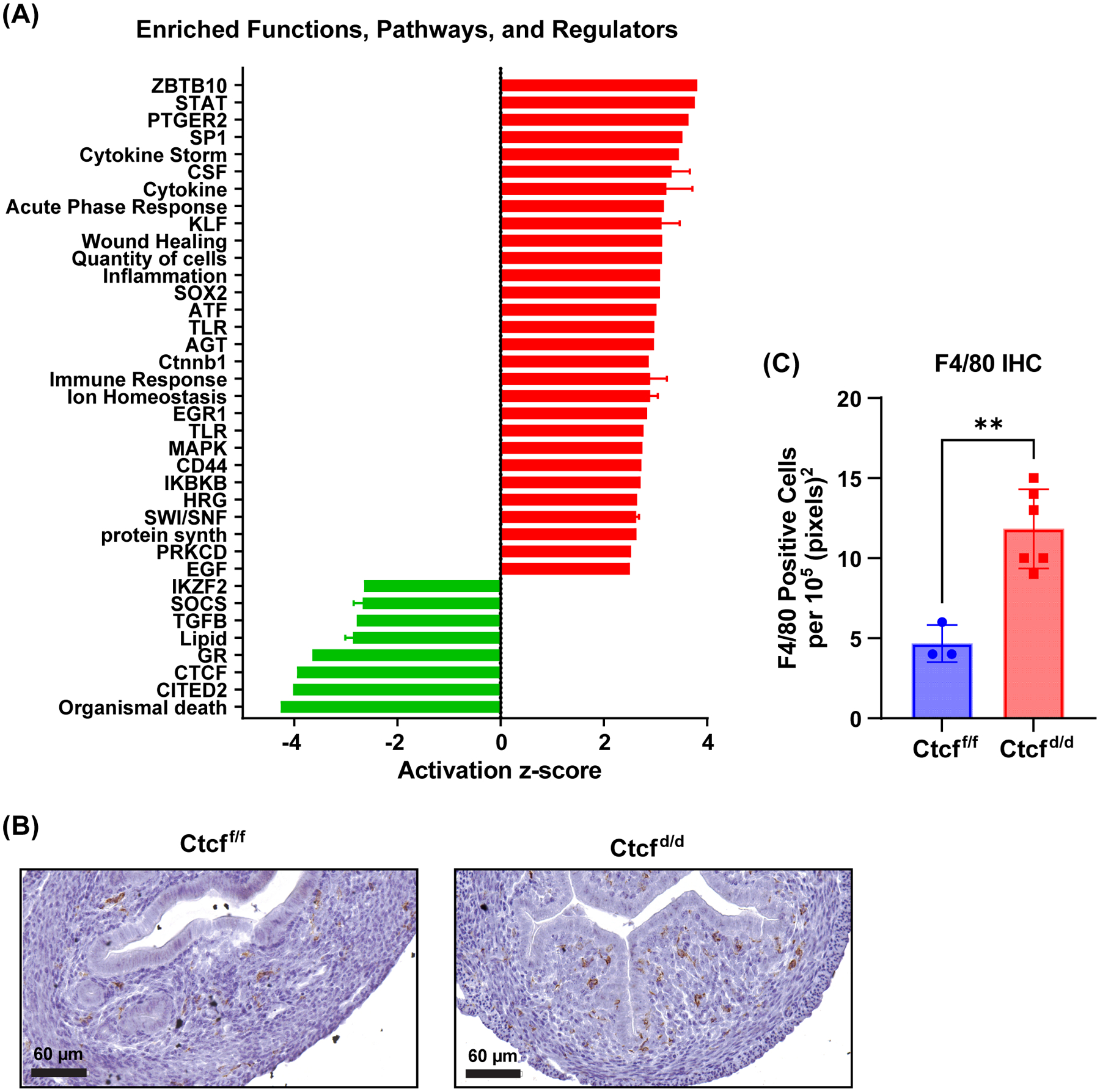
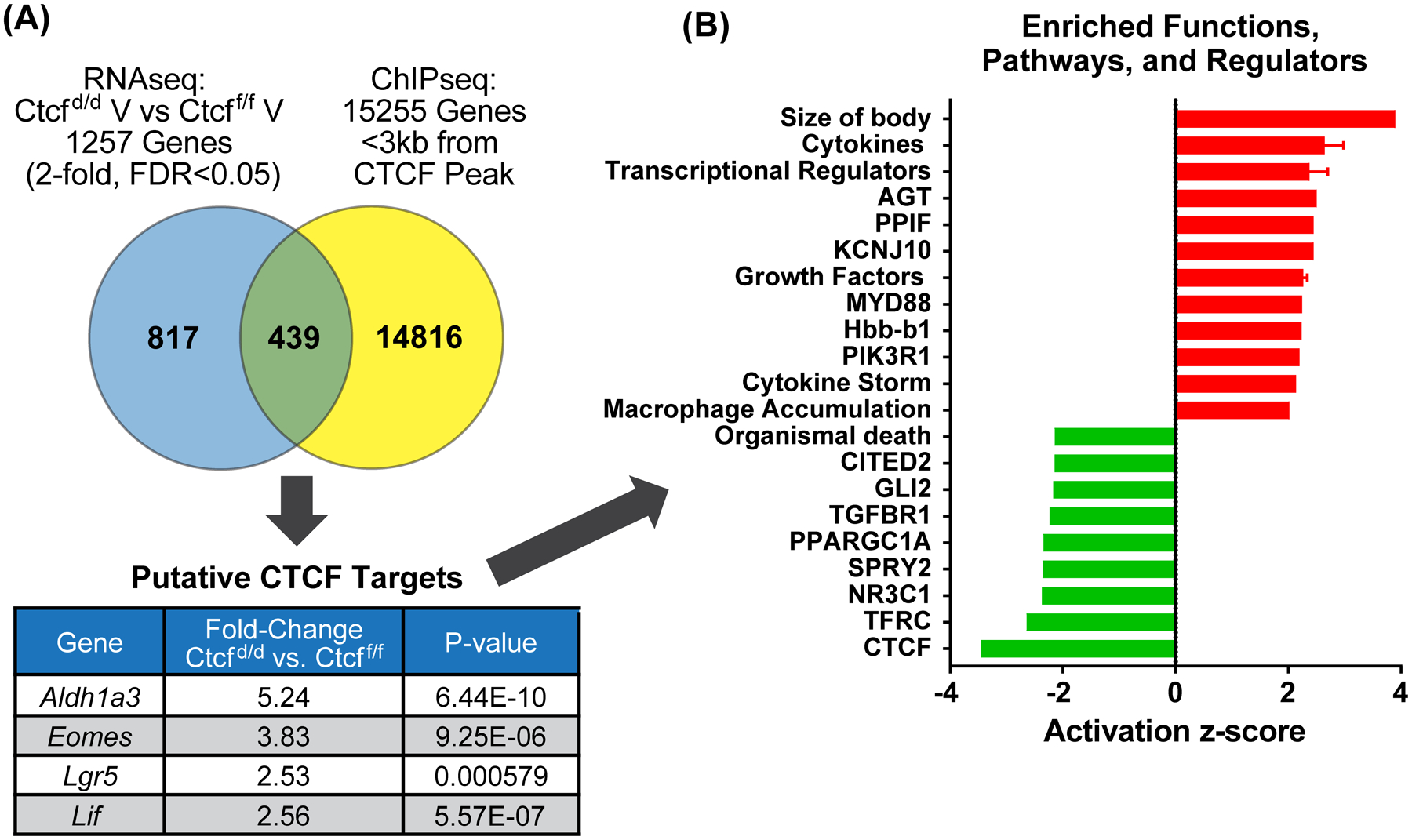
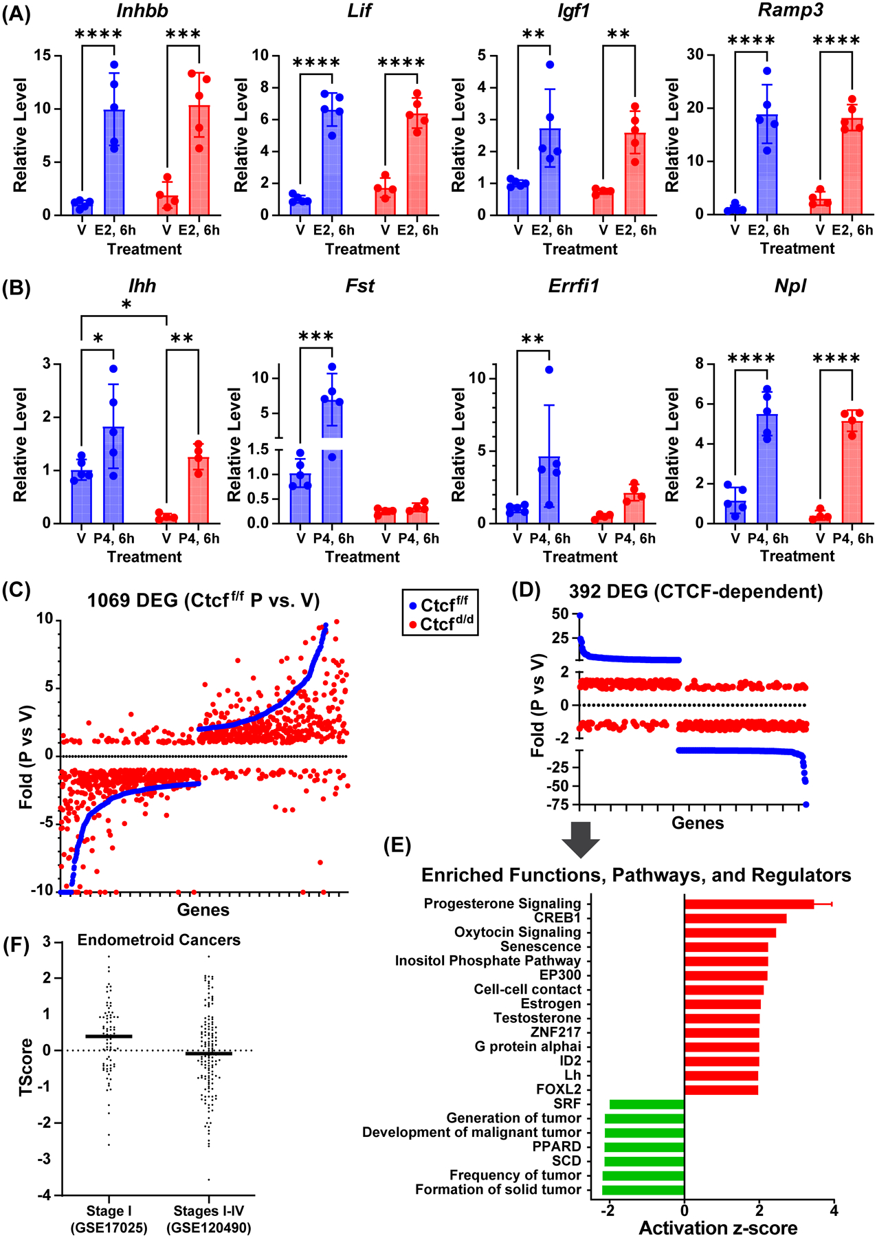
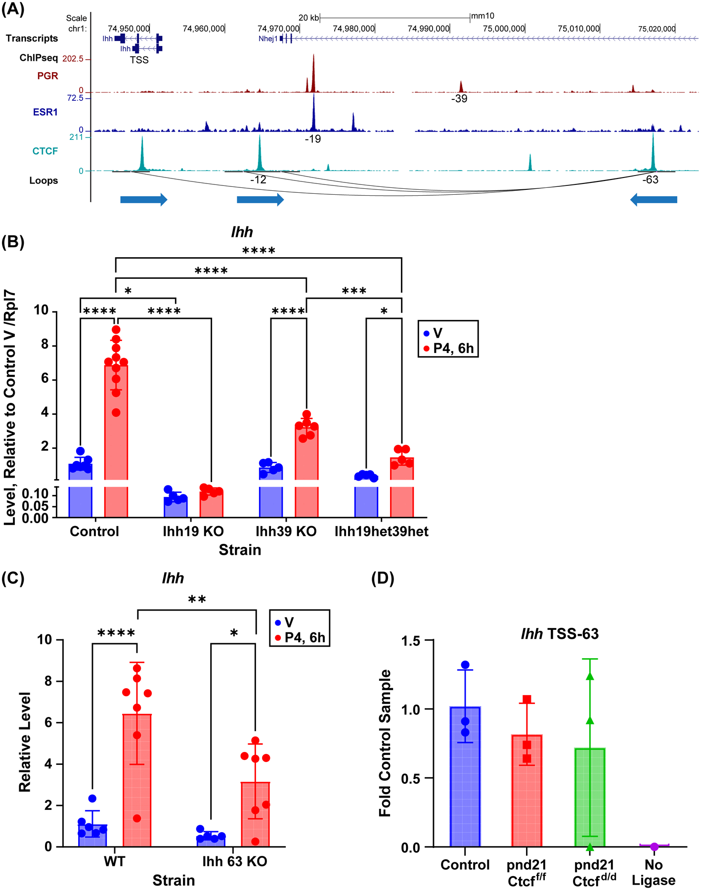
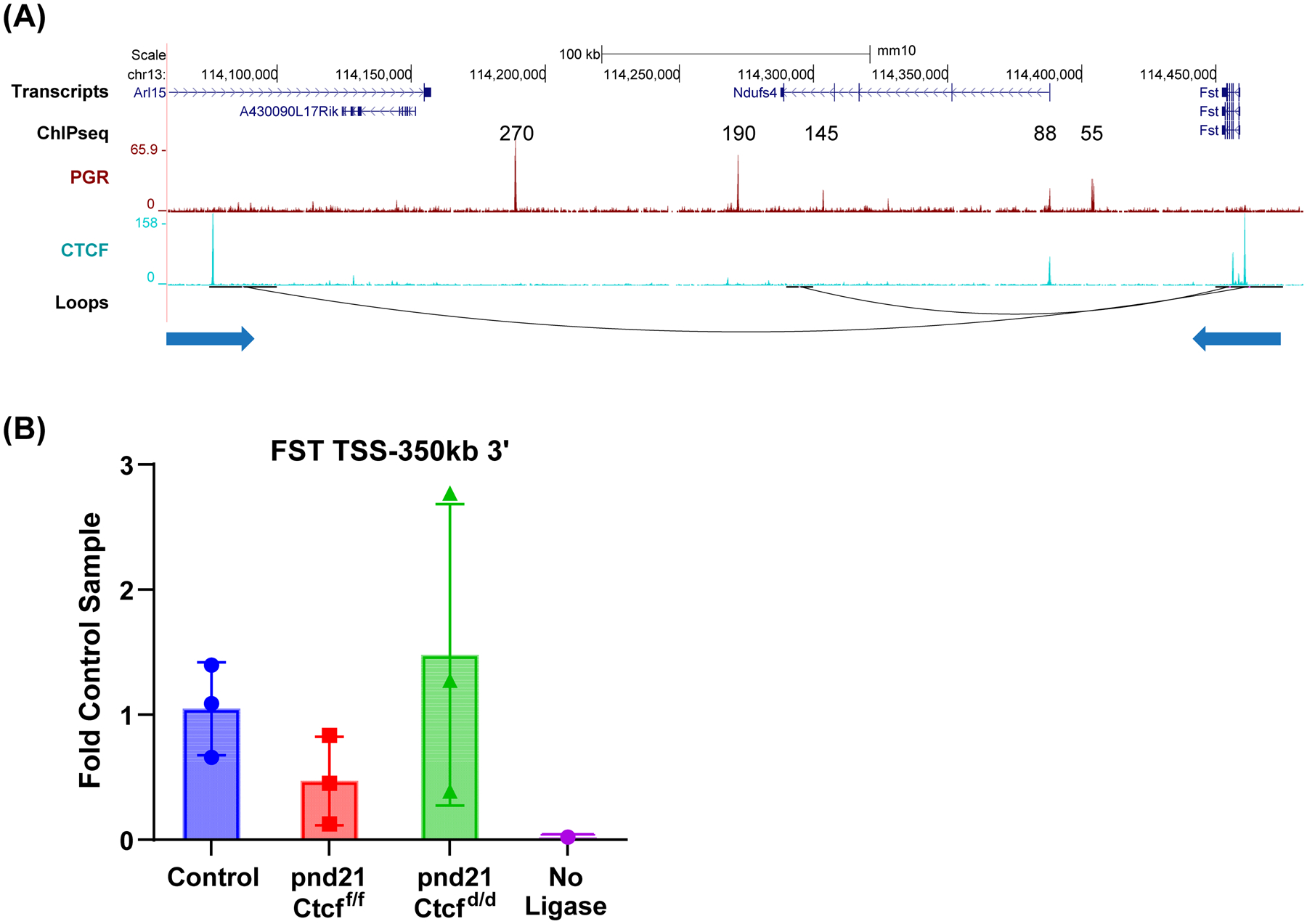
Receptors for estrogen and progesterone frequently interact, via Cohesin/CTCF loop extrusion, at enhancers distal from regulated genes. Loss-of-function CTCF mutation in >20% of human endometrial tumors indicates its importance in uterine homeostasis. To better understand how CTCF-mediated enhancer-gene interactions impact endometrial development and function, the Ctcf gene was selectively deleted in female reproductive tissues of mice. Prepubertal Ctcfd/d uterine tissue exhibited a marked reduction in the number of uterine glands compared to those without Ctcf deletion (Ctcff/f mice). Post-pubertal Ctcfd/d uteri were hypoplastic with significant reduction in both the amount of the endometrial stroma and number of glands. Transcriptional profiling revealed increased expression of stem cell molecules Lif, EOMES, and Lgr5, and enhanced inflammation pathways following Ctcf deletion. Analysis of the response of the uterus to steroid hormone stimulation showed that CTCF deletion affects a subset of progesterone-responsive genes. This finding indicates (1) Progesterone-mediated signaling remains functional following Ctcf deletion and (2) certain progesterone-regulated genes are sensitive to Ctcf deletion, suggesting they depend on gene-enhancer interactions that require CTCF. The progesterone-responsive genes altered by CTCF ablation included Ihh, Fst, and Errfi1. CTCF-dependent progesterone-responsive uterine genes enhance critical processes including anti-tumorigenesis, which is relevant to the known effectiveness of progesterone in inhibiting progression of early-stage endometrial tumors. Overall, our findings reveal that uterine Ctcf plays a key role in progesterone-dependent expression of uterine genes underlying optimal post-pubertal uterine development.

Keywords: chromatin; estrogen; progesterone; puberty; uterus.


Conflict of interest statement

Conflict of Interest Statement

The authors declare no conflicts of interest

Figures

Figure 1
Figure 1. Ctcf-null uteri develop post-pubertal hypoplasia
A. Uterine weights of uteri from Ctctf/f and Ctcfd/d mice (mg uterus normalized to g body weight); pre-pubertal (21 days) and post-pubertal (12 weeks and older). N=4 to 7 for each group. * p<0.05; ***p<0.001; ****p<0.0001 by 2 way ANOVA with uncorrected Fisher LSD post test. B. H&E stained uterine tissue sections from prepubertal (21-days old) and post-pubertal (6 weeks old) Ctcff/f and Ctcfd/d mice. Yellow arrows indicate stromal thickness. Scale bars indicate 60 or 200 μm. C. Stromal width and number of glands in uterine tissue. N=3 to 6 for each group. * p<0.05; p<0.001; by 2 way ANOVA with uncorrected Fisher LSD post test. D. IHC for CTCF protein in uterine sections from prepubertal (21 days) and post pubertal (6 weeks) Ctcff/f and Ctcfd/d mice. E. Analysis of Ki67 proliferative marker in 21-day old Ctcff/f and Ctcfd/d mice. Ki67 positive cells are brown. The % Ki67 positive cells were quantified in the luminal epithelium (epi) and the stroma (str) cells. **p<0.01; ****p<0.0001 by 2 way ANOVA with uncorrected Fisher LSD post test.
Figure 2
Figure 2. Transcriptional profile indicates prepubertal Ctcf deletion leads to an inflammatory response
A. Summary of functions and pathways enriched in prepubertal uterine Ctcfd/d vs. Ctcff/f gene set. Activation z-score indicates degree and direction of impact on the pathway (activation (red) or inhibition (green)). B. IHC for F4/80 reveals increased signal (brown) in uterine stroma of Ctcfd/d indicative of monocyte infiltration. C. F4/80 positive cells per 105 (pixels)2 n=3–5 t-test **p<0.01
Figure 3
Figure 3. Putative CTCF target genes include stem cell factors
A. Venn diagram comparing prepubertal uterine Ctcfd/d vs. Ctcff/f gene set to genes <3kb from a CTCF ChIPseq peak. Values for four stem cell genes are listed in table. B. Summary of functions and pathways enriched in the putative CTCF target gene set (439 genes).
Figure 4
Figure 4. Ctcf deficiency causes progesterone insensitivity in a subset of uterine genes
A. RT-PCR of estrogen responsive genes from prepubertal (21 days) Ctcff/f and Ctcfd/d uterine RNA. Mice were treated for 6 hours (6h) with sesame oil vehicle (V) or estrogen (E2). All genes were normalized to Rpl7. N=5 *p<0.05; **p<0.01; ***p<0.001; ****p<0.0001 by 2 way ANOVA with uncorrected Fisher LSD post test. B. RT-PCR of progesterone responsive genes from prepubertal (21 days) Ctcff/f and Ctcfd/d uterine RNA. Mice were treated for 6 hours (6h) with sesame oil vehicle (V) or progesterone (P4). All genes were normalized to RPL7. N=4–5 *p<0.05; **p<0.01; ***p<0.001; ****p<0.0001 by 2 way ANOVA with uncorrected Fisher LSD post test. C. Scatter plot of genes that are progesterone responsive in Ctcff/f uterus (blue) in rank order of fold change along X axis together with fold changes of same genes in Ctcfd/d uterine RNA samples (red). D. Scatter plot illustrating Ctcff/f progesterone responsive uterine genes (blue) that are progesterone insensitive in Ctcfd/d (red; absolute value fold change <1.5). E. Summary of functions and pathways enriched in Ctcff/f progesterone responsive uterine genes that lack response in Ctcfd/d. F. T-Scores calculated for CTCF-dependent progesterone target genes relative to transcriptomes from Stage I endometroid cancer (GSE17025) or from Stage I to Stage IV endometroid cancer (GSE120490).
Figure 5
Figure 5. Chromatin structures and hormone dependent enhancers mediate uterine Ihh regulation
A. PGR, ESR1 and CTCF ChIPseq and loops near Ihh gene. Both ESR1 and PGR interact with an enhancer 19 kb 5’ (–19) of the Ihh gene. PGR interacts with a second enhancer 39 kb 5’ of Ihh (–39). Interactions with Ihh occur via CTCF bound loop anchors at the Ihh gene and 63 kb 5’ (–63). An interaction between regions 12 kb 5’ of Ihh (–12) and the −63 region are also observed. The direction of teach CTCF motif is indicated by arrow under the loop anchors. B. RT-PCR of Ihh in ovariectomized adult uterine RNA from mice with deletion of enhancers −19 (Ihh19 KO) or −39 (Ihh39 KO), mice that are heterozygous for deletion of one copy each of the −19 and −39 enhancers (Ihh19het39het), and control littermates. Mice were treated for 6 hours (6h) with sesame oil vehicle (V) or progesterone (P4). N=5–10 *p<0.05; ***p<0.001; ****p<0.0001 by 2 way ANOVA with uncorrected Fisher LSD post test. C. RT-PCR of Ihh in ovariectomized adult uterine RNA from mice with deletion of a CTCF site at −63 (Ihh 63 KO) or their control littermates (WT). Mice were treated for 6 hours (6h) with sesame oil vehicle (V) or progesterone (P4). N=5–7 *p<0.05; **p<0.01; ****p<0.0001 by 2 way ANOVA with uncorrected Fisher LSD post test. D. 3C PCR to detect ligation between HindIII fragments at the Ihh TSS and the −63 CTCF binding loop anchor (IHH TSS-63) in adult mice (control) or prepubertal (pnd21) Ctcff/f and Ctcfd/d uterine DNA.
Figure 6
Figure 6. Chromatin structures and PGR binding enhancers interacting with Fst gene
A. PGR and CTCF ChIPseq and loops near the Fst gene. Interactions with Fst occur via CTCF bound loop anchors at the Fst gene and 350 kb 3’ of Fst in the Arl15 gene. The direction of each CTCF motif is indicated by the arrow underneath the loop anchor. B. 3C PCR to detect ligation between BamHI fragments at the Fst TSS and the 350kb 3’ CTCF binding loop anchor (FST TSS-350 3’) in adult mice (control) or prepubertal (pnd21) Ctcff/f and Ctcfd/d uterine DNA.

References

    1. Sikorska N, and Sexton T (2020) Defining Functionally Relevant Spatial Chromatin Domains: It is a TAD Complicated. Journal of Molecular Biology 432, 653–664 - PubMed
    1. DeMayo FJ, and Lydon JP (2020) 90 YEARS OF PROGESTERONE: New insights into progesterone receptor signaling in the endometrium required for embryo implantation. Journal of molecular endocrinology 65, T1–T14 - PMC - PubMed
    1. Hewitt SC, and Korach KS (2018) Estrogen Receptors: New Directions in the New Millennium. Endocrine reviews 39, 664–675 - PMC - PubMed
    1. Stewart CA, Fisher SJ, Wang Y, Stewart MD, Hewitt SC, Rodriguez KF, Korach KS, and Behringer RR (2011) Uterine gland formation in mice is a continuous process, requiring the ovary after puberty, but not after parturition. Biol Reprod 85, 954–964 - PMC - PubMed
    1. Hewitt SC, Carmona M, Foley KG, Donoghue LJ, Lierz SL, Winuthayanon W, and Korach KS (2020) Peri- and Postpubertal Estrogen Exposures of Female Mice Optimize Uterine Responses Later in Life. Endocrinology 161 - PMC - PubMed

Publication types