

Feasibility of [18F]fluoropivalate hybrid PET/MRI for imaging lower and higher grade glioma: a prospective first-in-patient pilot study
- PMID: 37490079
- PMCID: PMC10611885
- DOI: 10.1007/s00259-023-06330-0
Feasibility of [18F]fluoropivalate hybrid PET/MRI for imaging lower and higher grade glioma: a prospective first-in-patient pilot study
Abstract
Purpose: MRI and PET are used in neuro-oncology for the detection and characterisation of lesions for malignancy to target surgical biopsy and to plan surgical resections or stereotactic radiosurgery. The critical role of short-chain fatty acids (SCFAs) in brain tumour biology has come to the forefront. The non-metabolised SCFA radiotracer, [18F]fluoropivalate (FPIA), shows low background signal in most tissues except eliminating organs and has appropriate human dosimetry. Tumour uptake of the radiotracer is, however, unknown. We investigated the uptake characteristics of FPIA in this pilot PET/MRI study.
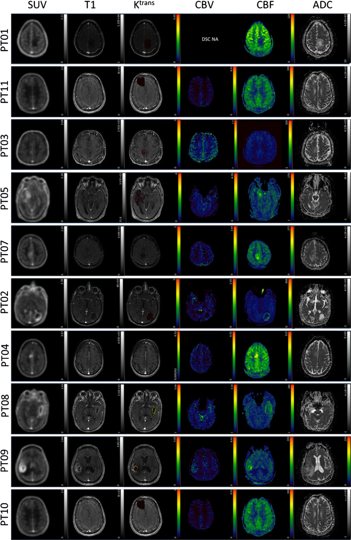
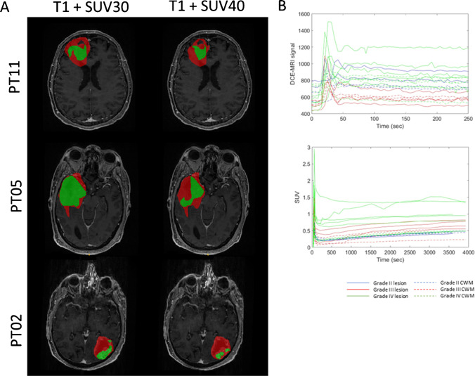
Methods: Ten adult glioma subjects were identified based on radiological features using standard-of-care MRI prior to any surgical intervention, with subsequent histopathological confirmation of glioma subtype and grade (lower-grade - LGG - and higher-grade - HGG - patients). FPIA was injected as an intravenous bolus injection (range 342-368 MBq), and dynamic PET and MRI data were acquired simultaneously over 66 min.
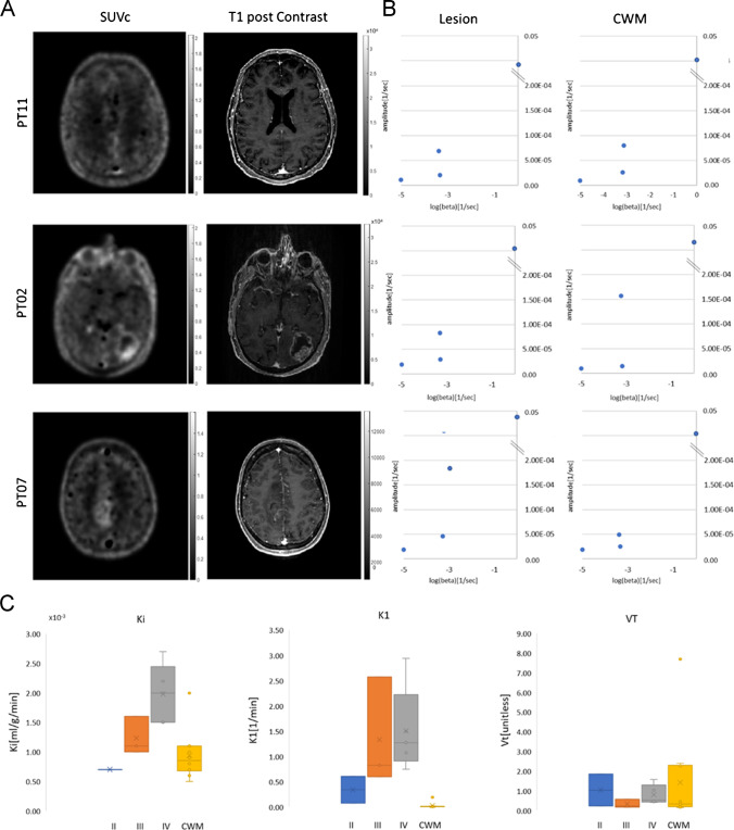
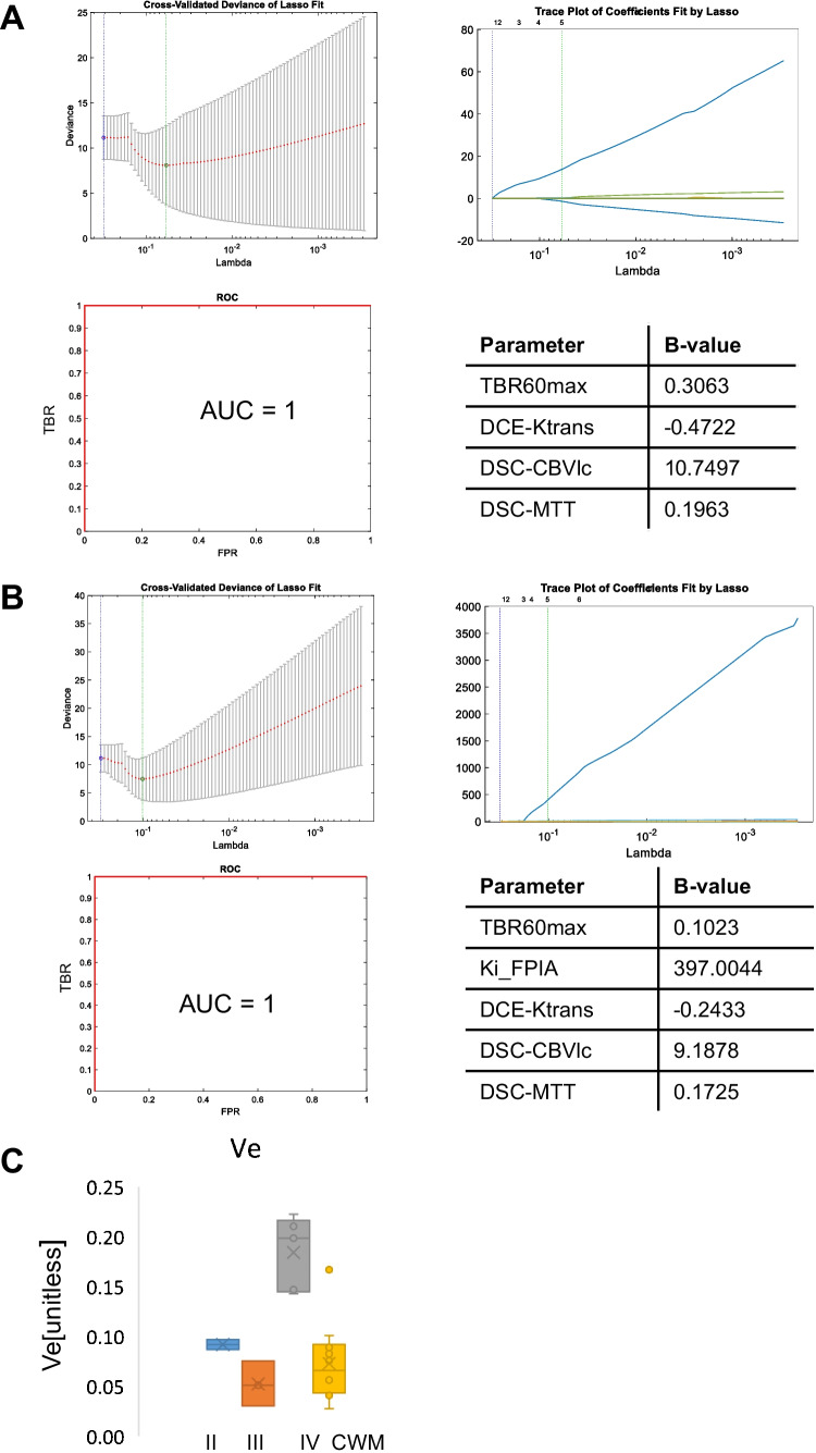
Results: All patients tolerated the PET/MRI protocol. Three patients were reclassified following resection and histology. Tumour maximum standardised uptake value (SUVmax,60) increased in the order LGG (WHO grade 2) < HGG (WHO grade 3) < HGG (WHO grade 4). The net irreversible solute transfer, Ki, and influx rate constant, K1, were significantly higher in HGG (p < 0.05). Of the MRI variables studied, DCE-MRI-derived extravascular-and-extracellular volume fraction (ve) was high in tumours of WHO grade 4 compared with other grades (p < 0.05). SLC25A20 protein expression was higher in HGG compared with LGG.
Conclusion: Tumoural FPIA PET uptake is higher in HGG compared to LGG. This study supports further investigation of FPIA PET/MRI for brain tumour imaging in a larger patient population.
Clinical trial registration: Clinicaltrials.gov, NCT04097535.
Keywords: FPIA; Glioma; PET/MRI; Short-chain fatty acid; [18F]fluoropivalate.
© 2023. The Author(s).
Conflict of interest statement
Financial interests: Eric O. Aboagye is inventor on a patent involving FPIA and has served in an advisor capacity to Radiopharm Theranostics Ltd and AstraZeneca. The authors declare no other relevant competing interests.
Figures
Comment in
-
[18F]Fluoropivalate, mitochondria, and the resurrection of short-chain fatty acids.Eur J Nucl Med Mol Imaging. 2023 Nov;50(13):3802-3805. doi: 10.1007/s00259-023-06367-1. Eur J Nucl Med Mol Imaging. 2023. PMID: 37523016 No abstract available.
Comment on
-
Clinical translation of 18F-fluoropivalate - a PET tracer for imaging short-chain fatty acid metabolism: safety, biodistribution, and dosimetry in fed and fasted healthy volunteers.Eur J Nucl Med Mol Imaging. 2020 Oct;47(11):2549-2561. doi: 10.1007/s00259-020-04724-y. Epub 2020 Mar 2. Eur J Nucl Med Mol Imaging. 2020. PMID: 32123971 Free PMC article.
References
Publication types
MeSH terms
Substances
Associated data
Grants and funding
LinkOut - more resources
Full Text Sources
Medical
