. 2023 Aug 3;127(30):6668-6674.
doi: 10.1021/acs.jpcb.3c04497. Epub 2023 Jul 25.

Spectroscopically Orthogonal Spin Labels in Structural Biology at Physiological Temperatures

Affiliations

Spectroscopically Orthogonal Spin Labels in Structural Biology at Physiological Temperatures

Markus Teucher et al. J Phys Chem B. .

Abstract

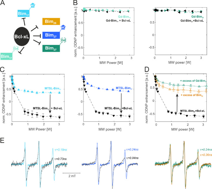
Electron paramagnetic resonance spectroscopy (EPR) is mostly used in structural biology in conjunction with pulsed dipolar spectroscopy (PDS) methods to monitor interspin distances in biomacromolecules at cryogenic temperatures both in vitro and in cells. In this context, spectroscopically orthogonal spin labels were shown to increase the information content that can be gained per sample. Here, we exploit the characteristic properties of gadolinium and nitroxide spin labels at physiological temperatures to study side chain dynamics via continuous wave (cw) EPR at X band, surface water dynamics via Overhauser dynamic nuclear polarization at X band and short-range distances via cw EPR at high fields. The presented approaches further increase the accessible information content on biomolecules tagged with orthogonal labels providing insights into molecular interactions and dynamic equilibria that are only revealed under physiological conditions.


Conflict of interest statement

The authors declare no competing financial interest.

Figures

Figure 1
Figure 1
DEER vs room temperature methods. Summary of the most relevant information, which can be extracted, and graphical sketch of the acquired data.
Figure 2
Figure 2
Minimal Bcl-2 interactome investigated. (A) Primary structures of Bim peptides used in this study with the position of the different spin labels highlighted. The spin labeling efficiency (η, spin/peptide concentration) is shown in parenthesis. A schematic color-coded representation of each spin-labeled peptide is shown on the right. The size of the peptides is encoded in the length of the colored boxes representing them. In the following, the Gd-Bim peptides will be named “Gd-peptides” for simplicity. (B) Ribbon representation of C-terminally truncated Bcl-xL (PDB: 4QVF) and (C) full length Bax (PDB: 1F16). Insets show a schematic representation of the unlabeled Bcl-xL used in this study and of the spin-labeled Bax carrying the nitroxide labels at the two natural cysteines 62 and 126.
Figure 3
Figure 3
Nitroxide kinetics in the presence and absence of Gd-peptides at physiological temperature. (A) Schematic representation of the interaction partners. (B) Cw EPR kinetics at 37 °C of nitroxide-labeled Bax in LUVs with a composition mimicking the outer mitochondrial membrane alone and with different Bim peptides. Bax was added at 20 μM final concentration, the peptides were added in a 1:1 stoichiometric ratio. (C) Cw EPR spectra extracted from the kinetic trace of panel B after 5 min (light gray) and 305 min (colored) incubation.
Figure 4
Figure 4
Room temperature ODNP on orthogonally labeled samples. (A) Schematic view of the probed interactions. (B) ODNP enhancement obtained at room temperature in aqueous solution in the presence of Gd-Bim peptides alone (final concentration 15 μM) or in the presence of Bcl-xL (1:1 stoichiometry). (C) ODNP enhancement obtained at room temperature in aqueous solution in the presence of MTSL-Bim peptides alone (final concentration 15 μM) or in the presence of Bcl-xL (1:1 stoichiometry). (D) Competition experiments performed with the MTSL-labeled peptides and Bcl-xL (1:1 stoichiometric ratio) upon addition of excess unlabeled or Gd-labeled Bim peptides in a 1:3 ratio with respect to the MTSL-peptide. Cw X-band EPR spectra detected on the ODNP samples presented above, respectively. Rotational correlation times were extracted using the approximation described in ref (35).
Figure 5
Figure 5
“Room temperature” (288 K) Gd–Gd distances by high field cw EPR. (A) Schematic representation of the interactions and cw EPR spectra of the central −1/2 to +1/2 transition detected on the Gd-Bim27 peptides in solution (final concentration 100 μM) in the absence or in the presence of Bcl-xL (1:1 stoichiometric ratio) or in the absence and presence of TFE (66% v/v). (B) Schematic of the interactions and cw EPR spectra on the Gd-Bim17 peptides under the same experimental conditions. The spectra detected at cryogenic temperatures are shown in Figure S4.

References

    1. Galazzo L.; Teucher M.; Bordignon E.. Orthogonal spin labeling and pulsed dipolar spectroscopy for protein studies. In Methods in Enzymology, Britt R. D. Ed.; Academic Press: 2022; Vol. 666, 79–119, 10.1016/bs.mie.2022.02.004. - DOI - PubMed
    1. Klose D.; Holla A.; Gmeiner C.; Nettels D.; Ritsch I.; Bross N.; Yulikov M.; Allain F. H.; Schuler B.; Jeschke G. Resolving distance variations by single-molecule FRET and EPR spectroscopy using rotamer libraries. Biophys. J. 2021, 120, 4842–4858. 10.1016/j.bpj.2021.09.021. - DOI - PMC - PubMed
    1. Peter M. F.; Gebhardt C.; Mächtel R.; Muñoz G. G. M.; Glaenzer J.; Narducci A.; Thomas G. H.; Cordes T.; Hagelueken G. Cross-validation of distance measurements in proteins by PELDOR/DEER and single-molecule FRET. Nat. Commun. 2022, 13, 4396.10.1038/s41467-022-31945-6. - DOI - PMC - PubMed
    1. Lueders P.; Jeschke G.; Yulikov M. Double Electron–Electron Resonance Measured Between Gd3+ Ions and Nitroxide Radicals. J. Phys. Chem. Lett. 2011, 2, 604–609. 10.1021/jz200073h. - DOI
    1. Kaminker I.; Yagi H.; Huber T.; Feintuch A.; Otting G.; Goldfarb D. Spectroscopic selection of distance measurements in a protein dimer with mixed nitroxide and Gd3+ spin labels. Phys. Chem. Chem. Phys. 2012, 14, 4355–4358. 10.1039/C2CP40219J. - DOI - PubMed

Publication types