. 2023 Aug 15;62(16):2461-2471.
doi: 10.1021/acs.biochem.3c00180. Epub 2023 Jul 25.

Introduction of Asymmetry in the Fused 4-Oxalocrotonate Tautomerases

Introduction of Asymmetry in the Fused 4-Oxalocrotonate Tautomerases

Kaci Erwin et al. Biochemistry. .

Abstract

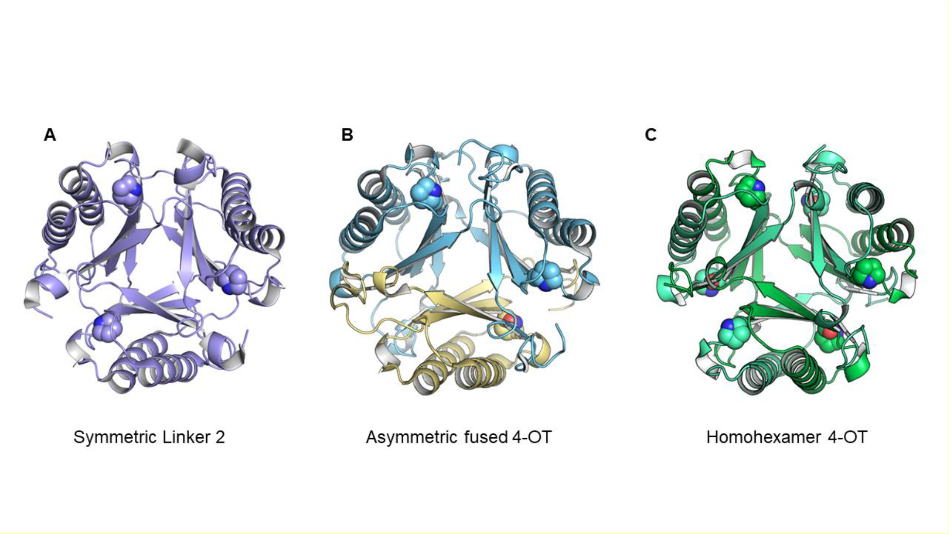
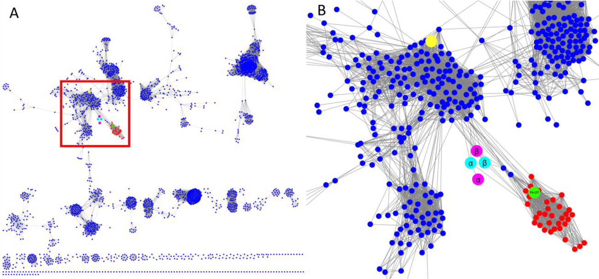
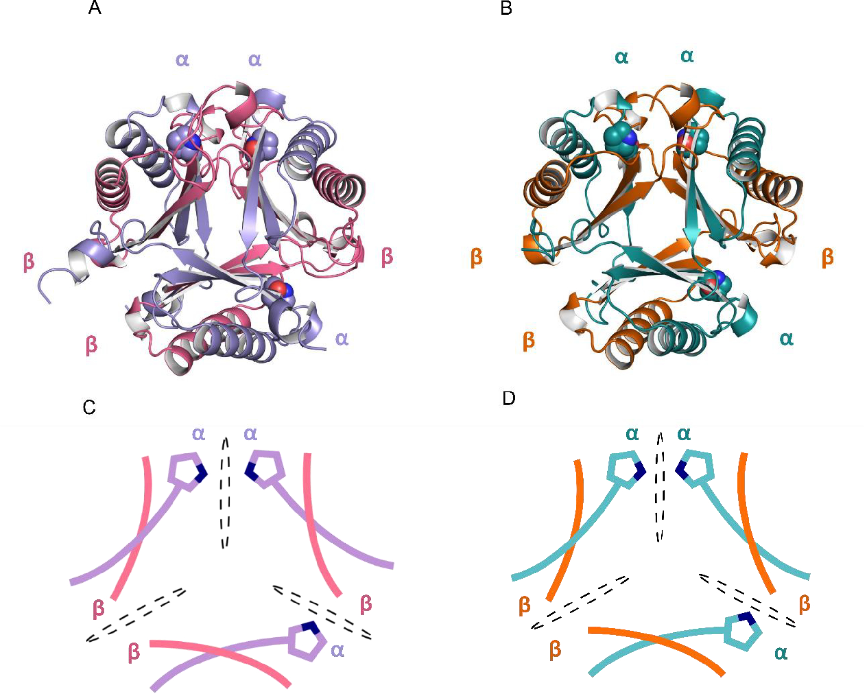
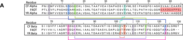
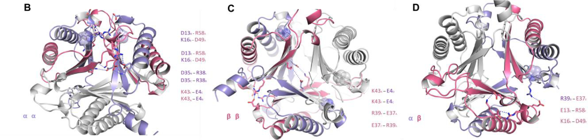
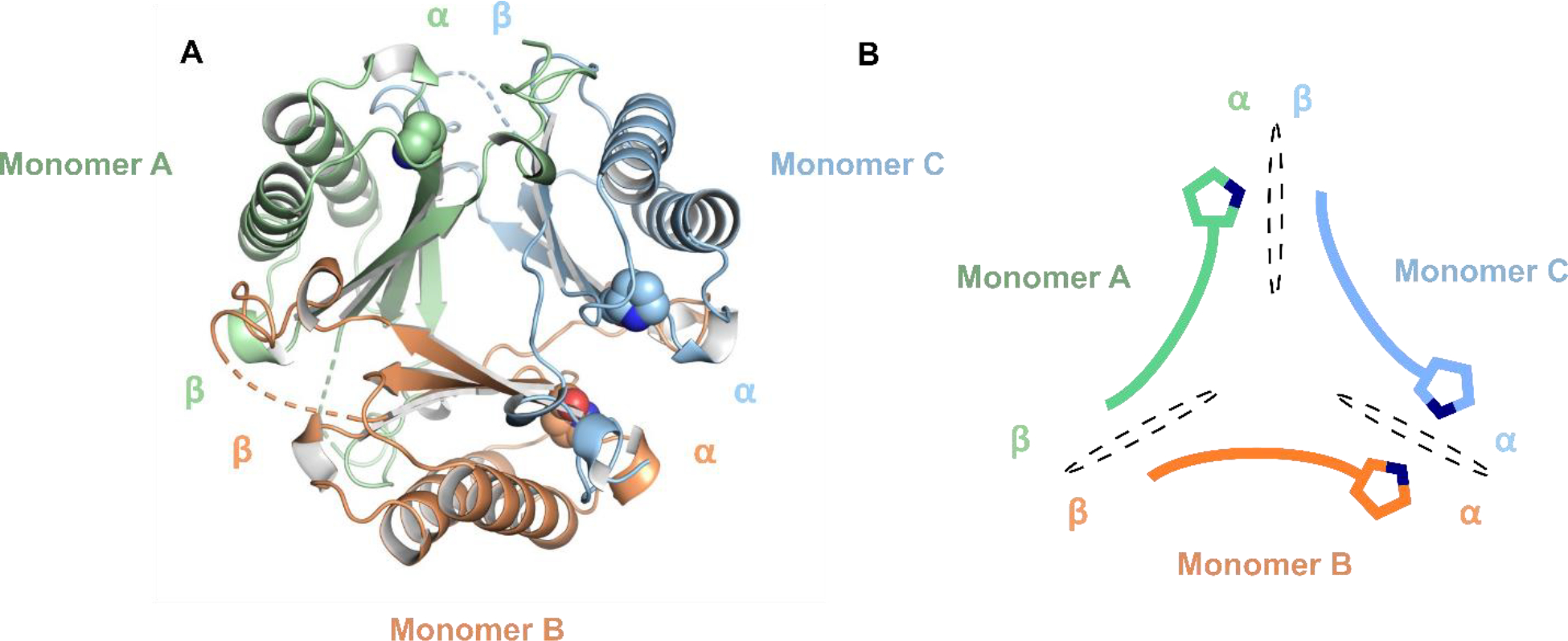
Members of the 4-oxalocrotonate tautomerase (4-OT) subgroup in the tautomerase superfamily (TSF) are constructed from a single β-α-β unit and form homo- or heterohexamers, whereas those of the other four subgroups are composed of two consecutively joined β-α-β units and form trimers. A subset of sequences, double the length of the short 4-OTs, is found in the 4-OT subgroup. These "fused" 4-OTs form a separate subgroup that connects to the short 4-OTs in a sequence similarity network (SSN). The fused gene can be a template for the other four subgroups, resulting in the diversification of activity. Analysis of the SSN shows that multiple nodes in the fused 4-OTs connect to five linker nodes, which in turn connect to the short 4-OTs. Some fused 4-OTs are symmetric trimers and others are asymmetric trimers. The origin of this asymmetry was investigated by subjecting the sequences in three linker nodes and a closely associated fourth node to kinetic, mutagenic, and structural analyses. The results show that each sequence corresponds to the α- or β-subunit of a heterohexamer that functions as a 4-OT. Mutagenesis indicates that the key residues in both are αPro1 and βArg-11, like that of a typical 4-OT. Crystallographic analysis shows that both heterohexamers are asymmetric, where one heterodimer is flipped 180° relative to the other two heterodimers. The fusion of two subunits (α and β) of one asymmetric heterohexamer generates an asymmetric trimer with 4-OT activity. Hence, asymmetry can be introduced at the heterohexamer level and then retained in the fused trimers.


Conflict of interest statement

The authors declare no competing financial interest.

Figures

Figure 1.
Figure 1.
Three representative oligomers in the TSF. (A) Representative symmetric trimer (PDB code 5UNQ)., (B) Representative asymmetric trimer (PDB code 6BLM)., (C) The canonical 4OT, a homohexamer (PDB code 1OTF). The catalytic proline residue is shown with a space-filling sphere.
Figure 2.
Figure 2.
The full level 2 network (90% representative network) for the (A) 4-OT subgroup and (B) blow-up of the region with the nodes of interest. (A) The level 2 network for the 4-OT subgroup where the region of interest is shown in the red box. (B) Detailed view of the nodes of interest. The nodes containing the short 4-OTs are shown in blue and those containing fused 4-OTs are shown in red. The node shown in green contains a fused 4-OT from Burkholderia lata (ATCC 17760, UniProt accession: Q392K7), which is an asymmetric trimer., The two putative heterohexamer 4-OTs are shown in cyan and magenta, where the subunits for the YR and CF heterohexamers are shown respectively in cyan and magenta. Each subunit has one node. The large yellow node at the top contains the 62-amino acid sequence for the canonical 4-OT.
Figure 3.
Figure 3.
Asymmetric heterohexamers YR- and CF-4OT (A) Ribbon diagram of Herbaspirillum sp. hhYR-4OT. The α- and β-domains are labeled and colored in purple and pink. (B) Ribbon diagram of Herbaspirillum sp. hhCF-4OT. The α-domain is colored in teal and the β-domain is colored in orange. (C-D) Schematic representation of the asymmetric arrangement of the two heterohexamers. Asymmetry is defined by three unique interfaces and are labeled as αα, αβ, and ββ. Each interface is divided by a dotted black oval. Pro1 is shown as a space-filling sphere in the cyan and gray at each interface. The color scheme is identical to that in A and B.
Figure 4.
Figure 4.
Conservation between hhYR/hhCF and fused 4OT, a representative asymmetric 4-OT trimer. (A) Multiple sequence alignment between the α- and β-chains of hhYR/hhCF and one protomer of the representative fused 4OT. Conserved salt bridge partners are highlighted and outlined between the α and β subunits. Highlighted in red is the linker region in fused 4OT Alignment was conducted with Clustal Omega. (B) Ribbon diagram of the salt bridge interactions in the ββ interface. Participating subunits are colored in purple (α-subunit) and pink (β-subunit) while chains that are not within the interface are in gray. Salt bridge interactions are shown with dotted blue lines and residues in stick form. Pro1 is shown as a sphere. (C) Salt bridge interactions within the αα interface. (D) Salt bridge interactions within the αβ interface.
Figure 4.
Figure 4.
Conservation between hhYR/hhCF and fused 4OT, a representative asymmetric 4-OT trimer. (A) Multiple sequence alignment between the α- and β-chains of hhYR/hhCF and one protomer of the representative fused 4OT. Conserved salt bridge partners are highlighted and outlined between the α and β subunits. Highlighted in red is the linker region in fused 4OT Alignment was conducted with Clustal Omega. (B) Ribbon diagram of the salt bridge interactions in the ββ interface. Participating subunits are colored in purple (α-subunit) and pink (β-subunit) while chains that are not within the interface are in gray. Salt bridge interactions are shown with dotted blue lines and residues in stick form. Pro1 is shown as a sphere. (C) Salt bridge interactions within the αα interface. (D) Salt bridge interactions within the αβ interface.
Figure 5.
Figure 5.
X-ray crystal structure of the asymmetric fYR trimer. (A) Ribbon diagram of the fused trimer determined at 2.3 Å resolution where each protomer is denoted by distinct colors. Pro1 is shown as space-filling spheres for each protomer. (B) Schematic representation of each protomer within the asymmetric fused trimer. Each interface is shown a black dotted oval.
Scheme 1.
Scheme 1.
Enzyme-catalyzed Tautomerization Reactions

References

    1. Davidson R, Baas B-J, Akiva E, Holliday G, Polacco BJ, LeVieux JA, Pullara CR, Zhang YJ, Whitman CP, and Babbitt PC (2018) A global view of structure-function relationships in the tautomerase superfamily. J. Biol. Chem. 293, 2342–2357. - PMC - PubMed
    1. Murzin AG (1996) Structural classification of proteins: new superfamilies. Curr. Opin. Struct. Biol. 6, 386–394. - PubMed
    1. Poelarends GJ, Veetil VP, and Whitman CP (2008) The chemical versatility of the β-α-β fold: Catalytic promiscuity and divergent evolution in the tautomerase superfamily. Cell. Mol. Life Sci. 65, 3606–3618. - PMC - PubMed
    1. LeVieux JA, Baas B-J, Kaoud TS, Davidson R, Babbitt PC, Zhang YJ, Whitman CP (2017) Kinetic and structural characterization of a cis-3-chloroacrylic acid dehalogenase homologue in Pseudomonas sp. UW4: A potential step between subgroups in the tautomerase superfamily. Arch Biochem Biophys, 636, 50–56. - PMC - PubMed
    1. Baas B-J, Medellin BP, LeVieux JA de Ruijter M, Zhang YJ, Brown SD, Akiva E, Babbitt PC, and Whitman CP (2019) Structural, kinetic, and mechanistic analysis of an asymmetric 4-oxalocrotonate tautomerase trimer. Biochemistry 58, 2617–2627. - PMC - PubMed

Publication types