. 2023 Jul 25;21(1):271.
doi: 10.1186/s12916-023-02982-9.

Assessment of bidirectional relationships between brain imaging-derived phenotypes and stroke: a Mendelian randomization study

Affiliations

Assessment of bidirectional relationships between brain imaging-derived phenotypes and stroke: a Mendelian randomization study

Ke Yu et al. BMC Med. .

Abstract

Background: Stroke is a major cause of mortality and long-term disability worldwide. Whether the associations between brain imaging-derived phenotypes (IDPs) and stroke are causal is uncertain.

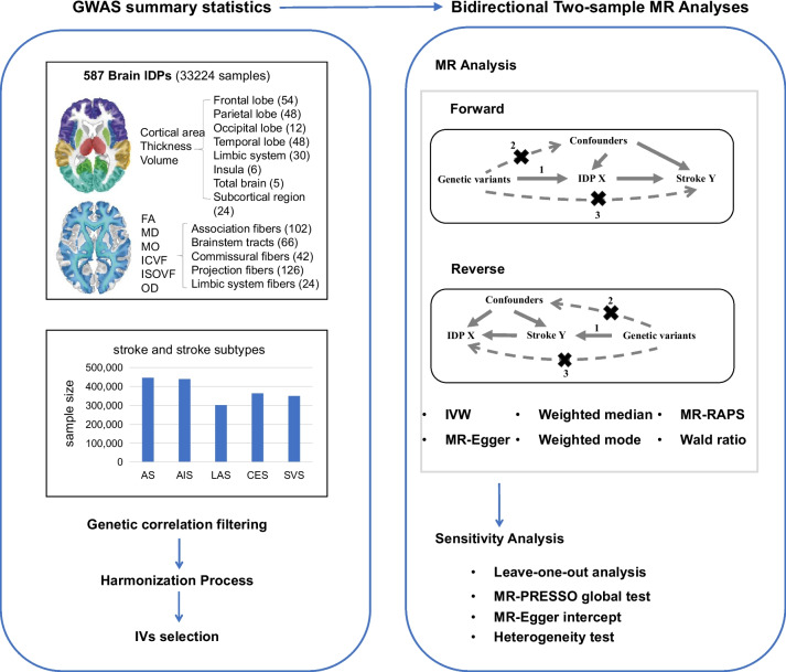
Methods: We performed two-sample bidirectional Mendelian randomization (MR) analyses to explore the causal associations between IDPs and stroke. Summary data of 587 brain IDPs (up to 33,224 individuals) from the UK Biobank and five stroke types (sample size range from 301,663 to 446,696, case number range from 5,386 to 40,585) from the MEGASTROKE consortium were used.

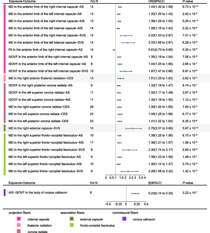
Results: Forward MR indicated 14 IDPs belong to projection fibers or association fibers were associated with stroke. For example, higher genetically determined mean diffusivity (MD) in the right external capsule was causally associated with an increased risk of small vessel stroke (IVW OR = 2.76, 95% CI 2.07 to 3.68, P = 5.87 × 10⁻¹²). Reverse MR indicated that genetically determined higher risk of any ischemic stroke was associated with increased isotropic or free water volume fraction (ISOVF) in body of corpus callosum (IVW β = 0.23, 95% CI 0.14 to 0.33, P = 3.22 × 10⁻⁷). This IDP is a commissural fiber and it is not included in the IDPs identified by forward MR.

Conclusions: We identified 14 IDPs with statistically significant evidence of causal effects on stroke or stroke subtypes. We also identified potential causal effects of stroke on one IDP of commissural fiber. These findings might guide further work toward identifying preventative strategies at the brain imaging levels.

Keywords: Causal association; IDPs; Mendelian randomization; Stroke.


Conflict of interest statement

The authors declare that they have no competing interests.

Figures

Fig. 1
Fig. 1
Workflow of the causal inference between IDPs and stroke. AS, any stroke; AIS, any ischemic stroke; CES, cardioembolic stroke; LAS, large artery stroke; MAF, minor allele frequency; LD, linkage disequilibrium; SVS, small vessel stroke
Fig. 2
Fig. 2
Causal associations between IDPs and stroke in the forward MR and reverse MR. The heatmap plot shows the causal associations between IDPs with at least one significant MR signals in forward and reverse MR and stroke. The pattern diagram in the bottom shows the brain anatomical region of corresponding IDPs. AS, any stroke; AIS, any ischemic stroke; CES, cardioembolic stroke; FA, fractional anisotropy; ISOVF, isotropic volume fraction; MD, mean diffusivity; SVS, small vessel stroke
Fig. 3
Fig. 3
Causal associations in the forward and reverse MR using the MR-IVW method. The forest plot shows the significant causal associations with P value < 5.91 × 10⁻⁵ and the estimated OR (or beta) with 95% confidence intervals (CI). AS, any stroke; AIS, any ischemic stroke; CES, cardioembolic stroke; FA, fractional anisotropy; ISOVF, isotropic volume fraction; MD, mean diffusivity; SVS, small vessel stroke

References

    1. Roth GA, et al. Global burden of cardiovascular diseases and risk factors, 1990–2019: update from the GBD 2019 study. J Am Coll Cardiol. 2020;76(25):2982–3021. doi: 10.1016/j.jacc.2020.11.010. - DOI - PMC - PubMed
    1. Bardutzky J, et al. Characterizing tissue fate after transient cerebral ischemia of varying duration using quantitative diffusion and perfusion imaging. Stroke. 2007;38(4):1336–44. doi: 10.1161/01.STR.0000259636.26950.3b. - DOI - PMC - PubMed
    1. Evans TE, et al. White matter microstructure improves stroke risk prediction in the general population. Stroke. 2016;47(11):2756–2762. doi: 10.1161/STROKEAHA.116.014651. - DOI - PubMed
    1. Debette S, Markus HS. The clinical importance of white matter hyperintensities on brain magnetic resonance imaging: systematic review and meta-analysis. Bmj. 2010;341:c3666. doi: 10.1136/bmj.c3666. - DOI - PMC - PubMed
    1. Buyck JF, et al. Cerebral white matter lesions are associated with the risk of stroke but not with other vascular events: the 3-City Dijon Study. Stroke. 2009;40(7):2327–31. doi: 10.1161/STROKEAHA.109.548222. - DOI - PubMed

Publication types