. 2023 Jul 26;8(1):279.
doi: 10.1038/s41392-023-01497-2.

Interaction between neutrophil extracellular traps and cardiomyocytes contributes to atrial fibrillation progression

Affiliations

Interaction between neutrophil extracellular traps and cardiomyocytes contributes to atrial fibrillation progression

Li He et al. Signal Transduct Target Ther. .

Abstract

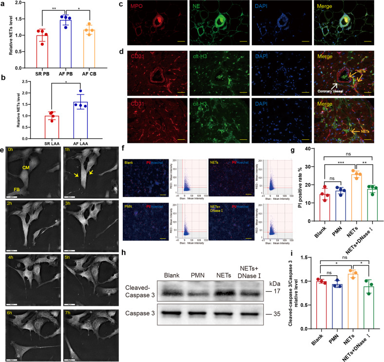
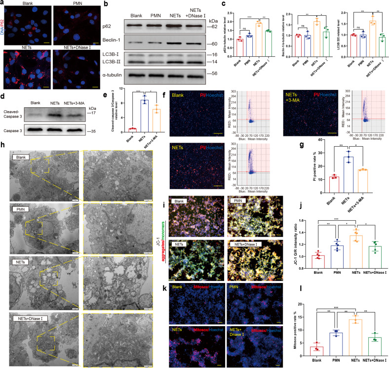
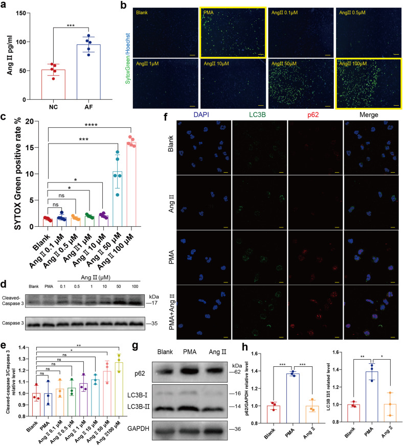
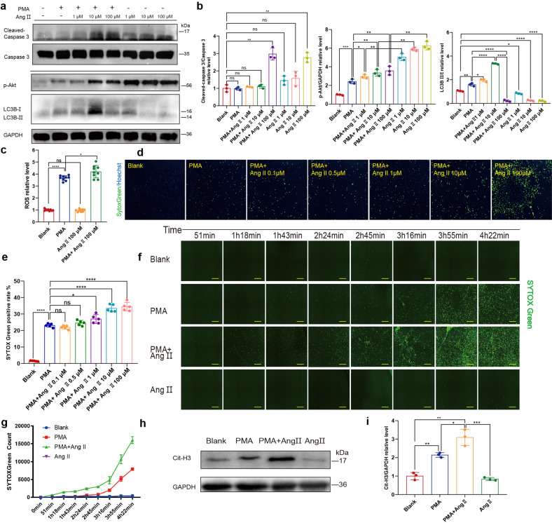
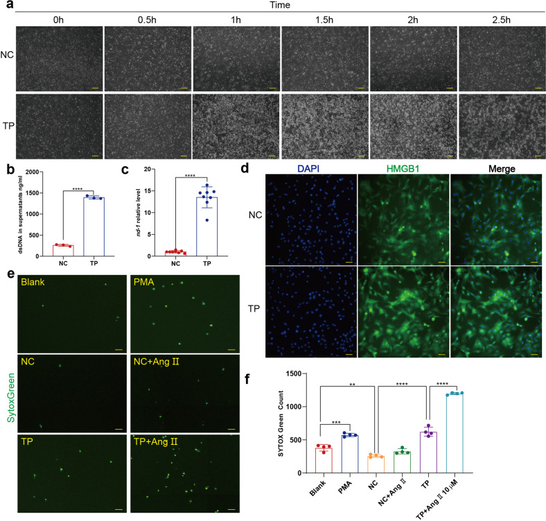
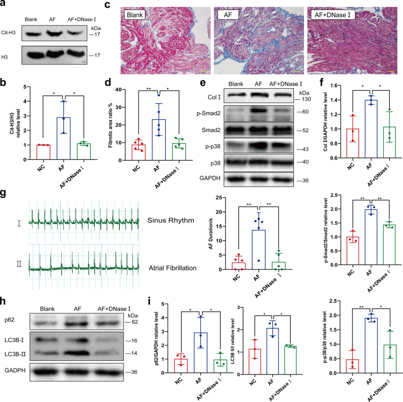
Atrial fibrillation (AF) is a frequent arrhythmia associated with cardiovascular morbidity and mortality. Neutrophil extracellular traps (NETs) are DNA fragments with cytoplasm proteins released from neutrophils, which are involved in various cardiovascular diseases. To elucidate the role of NETs in AF, we investigated the effect of NETs on AF progression and the secretion of NETs in AF. Results showed that: NETs induced the autophagic apoptosis of cardiomyocytes, and NETs also led to mitochondrial injury by promoting mitochondrial depolarization and ROS production. Ongoing tachy-pacing led to the structural loss of cardiomyocytes and provided potent stimuli to induce NETs secretion from neutrophils. In the meanwhile, increased Ang II in AF facilitated NETs formation through the upregulation of AKT phosphorylation, while it could not directly initiate NETosis as the autophagy was not induced. In vivo, DNase I was administrated to abrogate NETs formation, and AF-related fibrosis was ameliorated as expected. Correspondingly, the duration of the induced AF was reduced. Our study addresses the formation mechanism of NETs in AF and demonstrates the lethal effects of NETs on cardiomyocytes through the induction of mitochondrial injury and autophagic cell death, which comprehensively describes the positive feedback comprised of NETs and stimuli secreted by cardiomyocytes that sustains the progression of AF and AF related fibrosis.


Conflict of interest statement

Dr Yingqiang Guo and other authors have no financial or personal relationship with individuals or institutions that would inappropriately influence this work.

Figures

Fig. 1
Fig. 1
NETs are increased in AF patients and cytotoxic to cardiomyocytes. a Complex enzyme-linked immunosorbent assay (ELISA) experiment comparing NETs in the peripheral blood and coronary blood of patients with AF or SR (n = 4). b Comparison of NETs concentration in left atrial appendage homogenates of patients with AF and SR (n = 4). c NETs formed in the coronary vessels of AF patients. LAA section was stained for DNA (blue), NE (green) and MPO (red). Scale bar: 100 μm. MPO and NE double positive structure was defined as NETs. d NETs formed outside the coronary vessels of AF patients. LAA section was stained for DNA (blue), cit-H3 (green) and CD31 (red). Scale bar: 100 μm. Coronary vessels were labelled with CD31 and NETs were identified as cit-H3 positive. e Cardiomyocytes incubated with NETs underwent atrophy and perinuclear granule increase. Yellow arrows indicate NETs binding to the surface of cardiomyocytes. Scale bar: 20 μm. f, g Death rate of cardiomyocytes measured by Celigo analysis in the presence or absence of NETs and supernatants of PMN (n = 4). Cells in 96well plate were stained for all DNA (blue) and nucleuses of dead cells (red). Scale bar: 150 μm. Hoechst single positive cell was defined as the living, while Hoechst and PI double-positive cell was defined as the dead. h, i Increased cleaved caspase3/caspase 3 ratio in cardiomyocytes incubated with NETs analyzed through WB (n = 3). SR sinus rhythm, AF atrial fibrillation, PB peripheral blood, LAA left atrial appendage, CB coronary blood, NE neutrophil elastase, MPO myeloperoxidase, PMN polymorphonuclear leukocytes, PI propidium iodide. *P < 0.05, **P < 0.01, ***P < 0.001, ns (not significant). Data were presented as mean ± SD
Fig. 2
Fig. 2
NETs induce the autophagic cell death and mitochondrial injury of cardiomyocytes through the upregulation of autophagy. a Increased p62 positive granules in cardiomyocytes incubated with NETs demonstrated by IF staining microscopy. Cells in 24-well plate on coverslips were stained for DNA (blue) and p62 (red). p62 positive granules were imaged with a confocal laser-scanning microscope. Scale bar: 10 μm. b, c In vitro p62, Beclin-1 and LC3B II/I were all upregulated in cardiomyocytes incubated with NETs (n = 3). d, e In vitro 3-MA partly compromised the upregulation of cleaved- caspase 3 in cardiomyocytes induced by NETs (n = 3). f, g Death rate of cardiomyocytes measured by Celigo analysis in the presence or absence of NETs and NETs+3MA (n = 3). Cells in 96 well plate were stained for all DNA (blue) and nucleuses of dead cells (red). Scale bar: 100 μm. Hoechst single-positive cell was defined as the living, while Hoechst and PI double-positive cell was defined as the dead. h In vitro mitochondrial morphology damage was induced by NETs in cardiomyocytes, including swelling and reduction of cristae. Mitochondria were imaged with transmission electron microscope. Scale bar: 1 μm or 2 μm. i, j In vitro mitochondrial membrane potential was decreased in cardiomyocytes incubated with NETs (n = 5). The mitochondrial membrane potential of cardiomyocytes in 6-well plate in the presence or absence of NETs, degraded NETs with DNase I and supernatants of PMN using JC-1 and Hoechst was imaged by fluorescence microscope. Scale bar: 50 μm. The mitochondrial membrane potential of cardiomyocytes in 96-well plate was measured by fluorescence microplate reader. k, l In vitro excessive mtROS was produced in cardiomyocytes incubated with NETs (n = 3). Cells in 6-well and 96-well plate were stained for DNA (blue) and mtROS (red). mtROS of cardiomyocytes in 6-well plate in the presence or absence of NETs, degraded NETs with DNase I and supernatants of PMN using mitoSOX and Hoechst was imaged by Fluorescence microscope. Scale bar: 50 μm. Cardiomyocytes in 96well plate of increased mtROS production was compared by Celigo analysis. PMN polymorphonuclear leukocytes, mtROS mitochondrial reactive oxygen species. PI propidium iodide. *P < 0.05, **P < 0.01, ***P < 0.001, ns (not significant). Data were presented as mean ± SD
Fig. 3
Fig. 3
Ang II could not initiate NETosis. a ELISA experiment comparing Ang II in peripheral blood of patients with AF and normal control (n = 5). b, c Neutrophils of cfDNA measured by Celigo analysis in the presence or absence of PMA and Ang II of different concentration (0.1 μM, 0.5 μM, 1 μM, 10 μM, 50 μM or 100 μM) (n = 5). Neutrophils deprived from peripheral blood of the donator in 96-well plate was stained for all DNA (blue) and cfDNA (green). Scale bar: 100 μm. Hoechst single positive cell was defined as the living, while Hoechst and SYTOX Green double positive cell was defined as the neutrophil with cfDNA. Representatives with yellow frame demonstrate that despite the increase of SYTOX Green positive rate induced by Ang II, no linear or reticular DNA structure was identified. d, e Apoptosis analysis of Neutrophils incubated with PMA or Ang II of different concentration (0.1 μM, 0.5 μM, 1 μM, 10 μM, 50 μM or 100 μM) by comparing cleaved caspase 3/ caspase 3 ratio determined through WB(n = 3). f In vitro autophagy was not upregulated in neutrophils incubated with Ang II alone. Neutrophils in 24-well plate on coverslips were stained for DNA (blue), LC3B (green) and p62 (red). p62 or LC3B positive subcellular structures were imaged with a confocal laser-scanning microscope. Scale bar: 10 μm. g, h Analysis of the effect of Ang II on autophagy in neutrophils by comparing p62 and LC3B II/I ratio determined through WB (n = 3). Ang II angiotensin II, PMA Phorbol 12-myristate 13-acetate. *P˂0.05, **P < 0.01, ***P < 0.001, ****P < 0.0001, ns (not significant). Data were presented as mean ± SD
Fig. 4
Fig. 4
Ang II promotes NETosis and NETs secretion induced by PMA. a, b Apoptosis and autophagy analysis of human PMNs treated with PMA, Ang II of different concentrations (1 μM, 10 μM and 100 μM) or PMA+ Ang II. Analysis of p-Akt and LC3B II/I ratio (NETosis-related signaling) of PMNs treated with PMA, Ang II of different concentrations (1 μM, 10 μM and 100 μM) or PMA+ Ang II of different concentrations (1 μM, 10 μM and 100 μM) through WB (n = 3). c In vitro Ang II further upregulated PMA-induced ROS production of PMNs (n = 9). Suspended neutrophils were incubated with PMA, Ang II or PMA+ Ang II in 1.5 ml Eppendorf tubes gently shaking, and added into 96-well plate for subsequent determination of ROS with DCHF-DA in fluorescence microplate reader. d, e Analysis of concentration-dependent effect of Ang II on NETs secretion induced by PMA determined by Celigo (n = 5). Rat neutrophils in 96-well plate treated with PMA, Ang II or PMA+ Ang II of different concentrations (0.1 μM, 0.5 μM, 1 μM, 10 μM and 100 μM) were stained for all DNA (blue) and cfDNA (green). Scale bar: 150 μm. Hoechst single positive cell was defined as the living, while Hoechst and SYTOX Green double positive cell with linear DNA structure formation was defined as cells undergoing NETosis. f, g Analysis of Time-dependent effect of 10 μM Ang II on NETs secretion induced by PMA determined by Celigo (n = 4). Scale bar: 1 mm. h, i Analysis of cit-H3 in neutrophils in the presence or absence of PMA, Ang II and PMA+ Ang II through WB (n = 3). Cit-H3, citrullinated histone 3. *P < 0.05, **P < 0.01, ***P < 0.001, ****P < 0.0001, ns (not significant). Data were presented as mean ± SD
Fig. 5
Fig. 5
Supernatants of tachy-paced cardiomyocytes could induce NETosis. a Neonatal rat cardiomyocyte could not survive from long period of tachy-pacing. Cardiomyocytes in 6-well plate paced by C-Pace EP Culture Stimulator of 1 Hz (NC) or 6 Hz (TP) were observed and imaged by microscopy every 30 minutes. Scale bar: 200 μm. b Analysis of dsDNA with PicoGreen in the supernatants of tachy-paced cardiomyocytes and normal control (n = 3). cfDNA in supernatants of tachy-paced cardiomyocytes and normal control were measured with PicoGreen incubated in 96-well plate by fluorescence microplate reader. c Analysis of mtDNA in the supernatants of tachy-paced cardiomyocytes and normal control (n = 8). d HMGB1 was translocated from nucleus to cytoplasm in tachy-paced cardiomyocytes. Cardiomyocytes in 6-well plate were stained for DNA (blue) and HMGB1 (green). Scale bar: 20 μm. e, f Rat neutrophils undergoing NETosis measured by Celigo analysis in the presence or absence of PMA, Ang II and supernatants of cardiomyocytes paced with frequency of 1 Hz or 6 Hz (n = 4). Rat neutrophils in 96-well plate were stained for all DNA (blue) and cfDNA (green). Hoechst single positive cell was defined as the living, while Hoechst and SYTOX Green double positive cell with decondensed nucleus was defined as cell undergoing NETosis. Scale bar: 500 μm. TP, tachy-pacing, HMGB1 high mobility group box 1. **P < 0.01, ***P < 0.001, ****P < 0.0001. Data were presented as mean ± SD
Fig. 6
Fig. 6
DNase I intravenous injection ameliorates AF-induced atrial fibrosis. a, b In vivo DNase I abrogated AF-related upregulation of cit-H3 (n = 3). Cit-H3 in left atrium of rats with or without pharmacal induction of AF and DNase I injection was compared through WB. c, d In vivo DNase I ameliorated AF-related fibrosis (n = 5). Fibrotic area of rat left atrium was analyzed with Masson’s Trichrome staining. Blue area was defined as the fibrotic, red area was defined as normal myocardium. Scale bar: 50 μm. e, f Analysis of Collagen I, p-p38 and p-smad2 in left atrium of rat with or without pharmacal induction of AF and DNase I injection determined through WB (n = 3). g In vivo DNase I ameliorate the prolonged AF duration by continual pharmacal induction of AF (n = 5). AF induced by Ach-CaCl2 in rats with or without continual pharmacal induction of AF and DNase I injection was recorded by ECG and AF duration was documented. h, i In vivo upregulated autophagy by pharmacal induction of AF could be partly compromised by DNase I injection (n = 3). Analysis of p62 and LC3B II/I ratio in the left atrium of rat with or without pharmacal induction of AF and DNase I injection determined through WB. ECG, electrocardiograph. *P < 0.05, **P < 0.01. Data were presented as mean ± SD

References

    1. Du X, et al. Atrial fibrillation prevalence, awareness and management in a nationwide survey of adults in China. Heart. 2021;107:535–541. doi: 10.1136/heartjnl-2020-317915. - DOI - PMC - PubMed
    1. Zhou ZQ, et al. [An epidemiological survey of atrial fibrillation in China] Zhonghua Nei Ke Za Zhi. 2004;43:491–494. - PubMed
    1. Benjamin EJ, et al. Independent risk factors for atrial fibrillation in a population-based cohort. The Framingham Heart Study. JAMA. 1994;271:840–844. doi: 10.1001/jama.1994.03510350050036. - DOI - PubMed
    1. Roberts JD, et al. Epigenetic Age and the Risk of Incident Atrial Fibrillation. Circulation. 2021;144:1899–1911. doi: 10.1161/CIRCULATIONAHA.121.056456. - DOI - PMC - PubMed
    1. January CT, et al. 2019 AHA/ACC/HRS Focused Update of the 2014 AHA/ACC/HRS Guideline for the Management of Patients With Atrial Fibrillation: A Report of the American College of Cardiology/American Heart Association Task Force on Clinical Practice Guidelines and the Heart Rhythm Society in Collaboration With the Society of Thoracic Surgeons. Circulation. 2019;140:e125–e151. doi: 10.1161/CIR.0000000000000665. - DOI - PubMed

Publication types