. 2023 Jul 25;14(1):4461.
doi: 10.1038/s41467-023-40186-0.

Ezh2 emerges as an epigenetic checkpoint regulator during monocyte differentiation limiting cardiac dysfunction post-MI

Affiliations

Ezh2 emerges as an epigenetic checkpoint regulator during monocyte differentiation limiting cardiac dysfunction post-MI

Julie Rondeaux et al. Nat Commun. .

Abstract

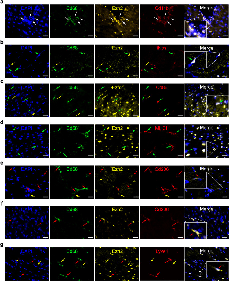
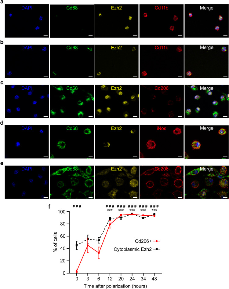
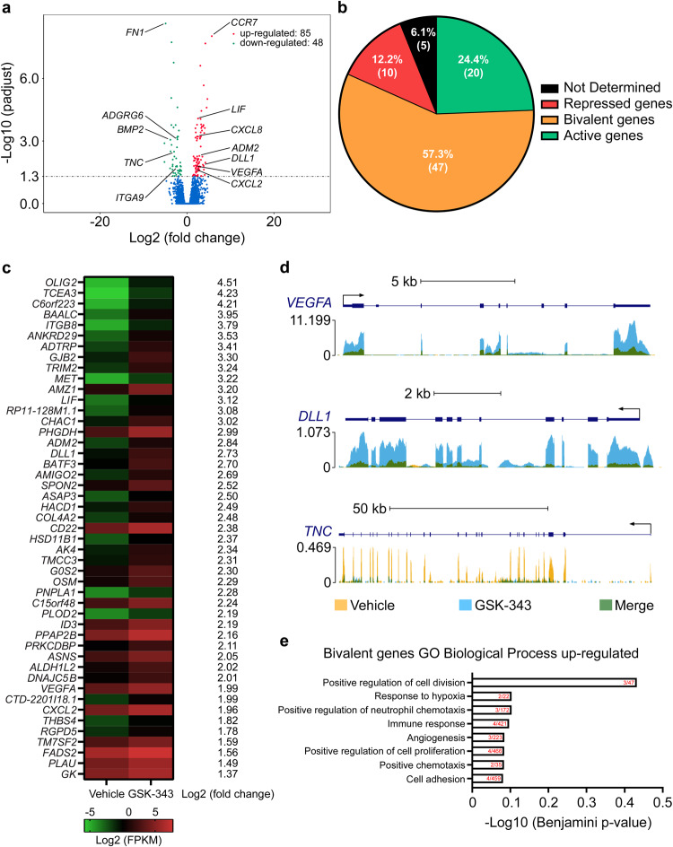
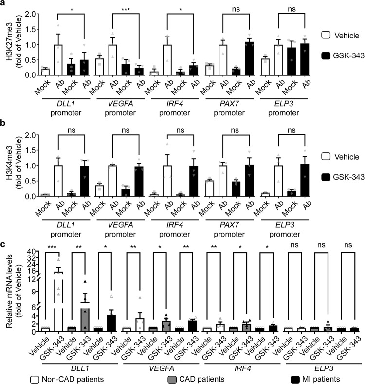
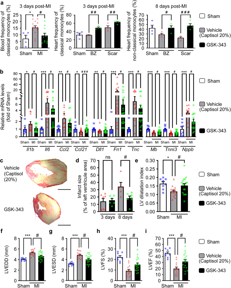
Epigenetic regulation of histone H3K27 methylation has recently emerged as a key step during alternative immunoregulatory M2-like macrophage polarization; known to impact cardiac repair after Myocardial Infarction (MI). We hypothesized that EZH2, responsible for H3K27 methylation, could act as an epigenetic checkpoint regulator during this process. We demonstrate for the first time an ectopic EZH2, and putative, cytoplasmic inactive localization of the epigenetic enzyme, during monocyte differentiation into M2 macrophages in vitro as well as in immunomodulatory cardiac macrophages in vivo in the post-MI acute inflammatory phase. Moreover, we show that pharmacological EZH2 inhibition, with GSK-343, resolves H3K27 methylation of bivalent gene promoters, thus enhancing their expression to promote human monocyte repair functions. In line with this protective effect, GSK-343 treatment accelerated cardiac inflammatory resolution preventing infarct expansion and subsequent cardiac dysfunction in female mice post-MI in vivo. In conclusion, our study reveals that pharmacological epigenetic modulation of cardiac-infiltrating immune cells may hold promise to limit adverse cardiac remodeling after MI.


Conflict of interest statement

The authors declare no competing interests.

Figures

Fig. 1
Fig. 1. Ezh2 is translocated to the cytoplasm in cardiac M2-like immunomodulatory macrophages in vivo.
Representative pictures of cardiac immunostaining for Ezh2 in a Cd11b+/Cd68- monocytes, b Cd68+/iNos+, c Cd68+/Cd86+, d Cd68+/MHCII+ pro-inflammatory differentiated macrophages, e Cd68+/Cd206+ immunomodulatory differentiated macrophages at 24 h after coronary ligation in mice to induce MI or f Cd68+/Cd206+, g Cd68+/Lyve1+ cardiac-resident immunomodulatory in healthy sham mice. Nuclei were stained with DAPI (blue), myeloid markers were Cd11b (red, panel a), Cd68 (green, panels ag), iNos (red, panel b), Cd86 (red, panel c), MHCII (red, panel d) Cd206 (red, panels e and f) and Lyve1 (red, panel g). Ezh2 (yellow) cellular localization was observed in each cell type but only appeared in immunomodulatory macrophages cytoplasm. Arrows indicate monocytes (white), non-determined macrophages (yellow), pro-inflammatory (green) and immunomodulatory macrophages (red), scale bars represent 25 μm. These cardiac immunostainings have been reproduced at least on three different mice with similar results.
Fig. 2
Fig. 2. Ezh2 is translocated to the cytoplasm in myeloid cells during M2 polarization in vitro.
Representative pictures of immunostaining for Ezh2 in a non-adherent monocytes, b adherent monocytes, c non-polarized M0 macrophages, d M1 macrophages and e M2 macrophages, differentiated and polarized in vitro. Nuclei were stained with DAPI (blue), monocytes were identified based on Cd11b (red, panels a and b) expression, and macrophages based on Cd68 (green, panels ae) expression. M1 or M2 macrophage phenotype was determined by expression of iNos (red, panel d) or Cd206 (red, panels c and e). Ezh2 (yellow) cellular localization was observed in each cell type. Scale bars represent 10 μm. These immunostainings have been reproduced at least 3 times from different mice with similar results. Kinetics of Ezh2 subcellular localization and Cd206 expression (f) were assessed by immunostaining during M2 macrophage polarization. Data are represented as percentage of total cells mean values ± SEM of 3 independent experiments (n = 3) each performed in duplicate for all time points. Asterisk (*) symbol indicates statistically significant difference between cells with either nuclear or cytoplasmic Ezh2 localization. Hashtag (#) depicts significant difference between Cd206 negative and positive cells. ### and ***p < 0.001; Two-way ANOVA with Sidak’s multiple comparisons test. Source data are provided as a Source Data file.
Fig. 3
Fig. 3. Ezh2-regulated bivalent genes in monocytes are implicated in cardiovascular repair processes.
Representative tracks and peak calling for promotors of inactive (PAX7), active (ELP3) and bivalent (DLL1, VEGFA, IRF4) genes (a) generated by ChIP-Seq analysis of human CD14+ monocytes. Representative Gene Ontology (GO) Biological Process categories significantly enriched for bivalent genes in human CD14+ monocytes. The bar graph represents the −log10 (Benjamini p-value), obtained from DAVID gene-enrichment in functional annotation terms after Fisher’s Exact test filled with the number of genes revealed by the analysis within the number of overall genes in each GO biological process category indicated in red (b). Quantification of H3K27me3 inactive (red) versus H3K4me3 active (green) histone marks in the promoter regions of three bivalent and two non-bivalent genes, assessed by ChIP-qPCR (c). Data represent mean fractions of input ± SEM of three independent experiments, corresponding to three different human donors, performed in duplicate (n = 3). Asterisk (*) symbol indicates H3K4me3 statistically significant enrichment compared to the inactive PAX7 promoter (Kruskal–Wallis test). Hashtag (#) depicts H3K27me3 significant enrichment compared to the active ELP3: *p < 0.05 and # p < 0.05. Source data are provided as a Source Data file.
Fig. 4
Fig. 4. Ezh2 pharmacological inhibition with GSK-343 increases bivalent gene expression and enhances human monocyte homing and angiogenic functions in vitro.
Global changes in gene expression upon GSK-343 treatment versus vehicle treatment analyzed by mRNA-seq (a) in cultured human monocytes. Volcano plot shows upregulated (red) and downregulated (green) genes (combined data from three different donors, n = 3) considered as statistically significant with a Benjamini-Hochberg adjusted p-value < 0.05. Classification of GSK-343-induced genes according to promoter status (b). Heatmaps of all significantly upregulated bivalent genes identified by RNA-seq analysis in human monocytes after GSK-343 treatment (c). Data obtained from three independent donors (n = 3) are expressed in log2 (FPKM). Representative tracks of changes induced by GSK-343 treatment on select up- and downregulated genes in human monocytes (d). Representative Gene Ontology (GO) Biological Process categories significantly (Benjamini-Hochberg adjusted p-value < 0.05) enriched for GSK-343-induced bivalent genes in human monocytes. The bar graph represents the −log10 (Benjamini-Hochberg adjusted p-value), obtained from DAVID gene-enrichment in functional annotation terms after Fisher’s Exact test filled with the number of genes revealed by the analysis within the number of overall genes in each GO biological process category indicated in red (e).
Fig. 5
Fig. 5. Pharmacological inhibition of EZH2 increases specifically bivalent gene expression through H3K27me3 demethylation in monocytes in vitro.
Enrichment of selected bivalent (DLL1, VEGFA, IRF4), inactive (PAX7) and active (ELP3) gene promoters in H3K27me3 (a) and H3K4me3 (b) as assessed by ChIP-qPCR. Ab indicates the enrichment obtain with either H3K27me3 (a) or H3K4me3 (b) targeting antibodies, while Mock represents the data obtain after immunoprecipitation with control IgG. Data are represented as mean fractions of input normalized to vehicle-treated monocytes ± SEM of three independent experiments corresponding to three different donors performed in duplicate (n = 3). The p values determined by Kruskal–Wallis test are depicted as asterisks in the figures as follows: ***p < 0.001; **p < 0.01; *p < 0.05; ns: non-significant enrichment compared to vehicle-treated monocytes. Transcript levels of indicated genes were measured by RT-qPCR following treatment with vehicle or GSK-343 of selected monocytes from non-coronary patients, CAD or AMI patients (c). RT-qPCR values are expressed as mean percentages of vehicle-treated monocytes ± SEM with B2M serving as internal control of four independent experiments corresponding to four different donors per group performed in duplicate (n = 4). The p values are depicted as asterisks after Kruskal–Wallis test in the figure as follows: ***p < 0.001; **p < 0.01; *p < 0.05; ns: non-significant. Source data are provided as a Source Data file.
Fig. 6
Fig. 6. Pharmacological Ezh2 inhibition with GSK-343 accelerates cardiac inflammatory resolution to prevent infarct expansion and subsequent cardiac remodeling and dysfunction post-MI.
Circulating (3 days post-MI) and cardiac (3 days and 8 days post-MI respectively for center and right panels) classical (Cd11bhiLy6chi) and non-classical (Cd11bhiLy6clo) monocyte population frequencies were evaluated by flow cytometry. Samples were collected from sham (n = 4) and MI mice treated daily either with vehicle (captisol 20%, n = 13 blood samples, n = 4 cardiac samples) or GSK-343 either from the infarcted scar area or from the healthy border zone (BZ). Data are expressed as mean frequency ± SEM of live CD45+/Cd19neg cells. b Cardiac transcript levels of indicated genes measured by RT-qPCR from sham (n = 8), or vehicle (n = 16) or GSK-343-treated (n = 18) mice at 8 days post-MI. Values expressed as mean percentages of sham ± SEM with B2m serving as internal control. c Representative pictures of infarct scar and M-mode echocardiography in sham, vehicle and GSK-343 treated mice at 8 days post-MI. Scale bar represents 1 mm. Infarct size assessed in vehicle (n = 7 and n = 5 respectively at 3 and 8 days post-MI) or GSK-343 treated (n = 6 and n = 7 respectively at 3 and 8 days post-MI) MI-mice (d). Data represent mean percentage of left ventricle area ± SEM. Quantitative analysis of LV dilatation index (e) and echo parameters for wall thickness: LVEDD (f), LVESD (g), LVFS (h) and LVEF (i) in sham (n = 8), vehicle (n = 14) and GSK-343 (n = 18) treated mice at 8 days post-MI. Data are presented as means ± SEM. For all panels (ai), asterisk (*) and hashtag (#) symbols indicates statistically significant difference compared to sham and vehicle condition respectively after Kruskal–Wallis test. The p values are depicted as follows: *** and ### p < 0.001, ** and ## p < 0.01, * and # p < 0.05; ns: non-significant. Source data are provided as a Source Data file.

References

    1. Swirski FK, Nahrendorf M. Cardioimmunology: the immune system in cardiac homeostasis and disease. Nat. Rev. Immunol. 2018;18:733–744. doi: 10.1038/s41577-018-0065-8. - DOI - PubMed
    1. Mouton AJ, et al. Mapping macrophage polarization over the myocardial infarction time continuum. Basic Res. Cardiol. 2018;113:26. doi: 10.1007/s00395-018-0686-x. - DOI - PMC - PubMed
    1. Leuschner F, et al. Rapid monocyte kinetics in acute myocardial infarction are sustained by extramedullary monocytopoiesis. J. Exp. Med. 2012;209:123–137. doi: 10.1084/jem.20111009. - DOI - PMC - PubMed
    1. van Amerongen MJ, Harmsen MC, van Rooijen N, Petersen AH, van Luyn MJ. Macrophage depletion impairs wound healing and increases left ventricular remodeling after myocardial injury in mice. Am. J. Pathol. 2007;170:818–829. doi: 10.2353/ajpath.2007.060547. - DOI - PMC - PubMed
    1. Panizzi P, et al. Impaired infarct healing in atherosclerotic mice with Ly-6C(hi) monocytosis. J. Am. Coll. Cardiol. 2010;55:1629–1638. doi: 10.1016/j.jacc.2009.08.089. - DOI - PMC - PubMed

Publication types