. 2023 Jul 25;14(1):4101.
doi: 10.1038/s41467-023-39586-z.

Macrophage-to-endothelial cell crosstalk by the cholesterol metabolite 27HC promotes atherosclerosis in male mice

Affiliations

Macrophage-to-endothelial cell crosstalk by the cholesterol metabolite 27HC promotes atherosclerosis in male mice

Liming Yu et al. Nat Commun. .

Abstract

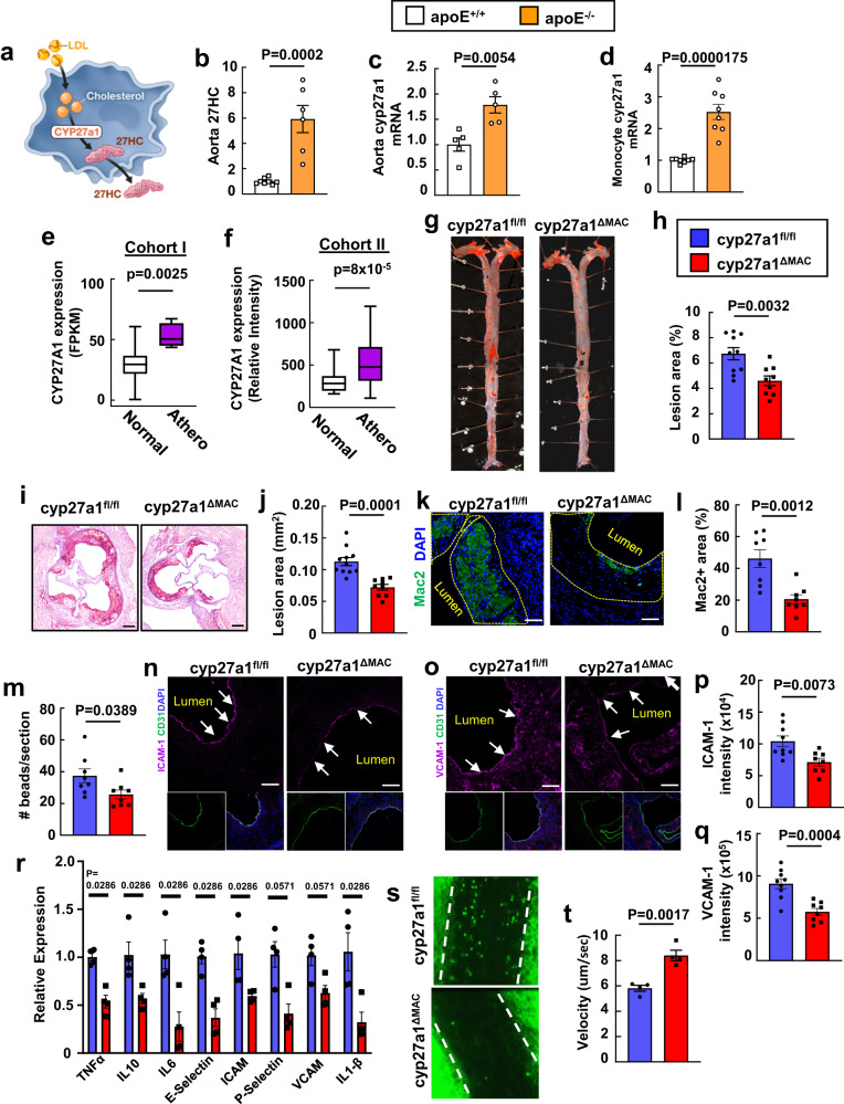
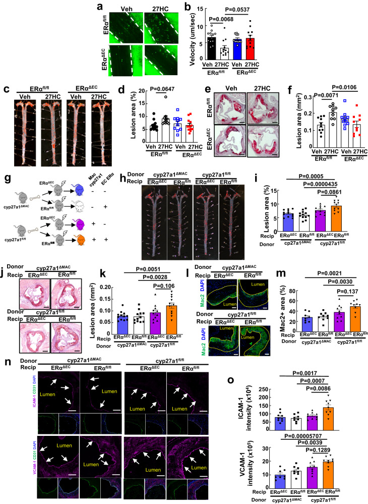
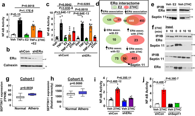
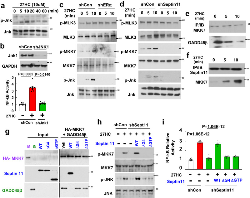
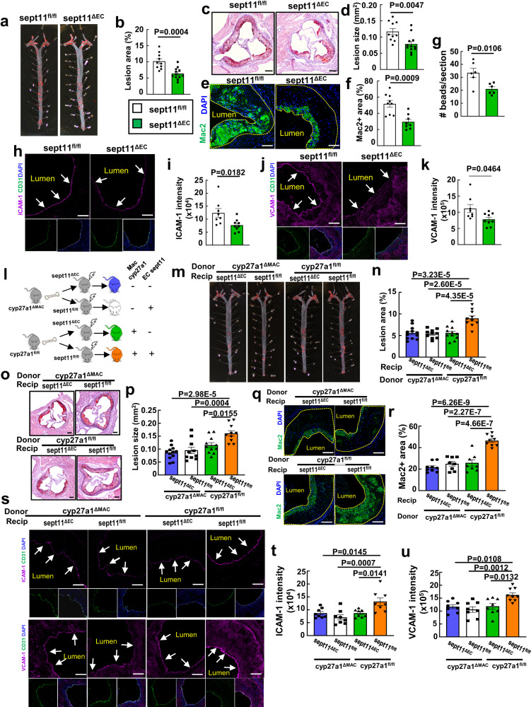
Hypercholesterolemia and vascular inflammation are key interconnected contributors to the pathogenesis of atherosclerosis. How hypercholesterolemia initiates vascular inflammation is poorly understood. Here we show in male mice that hypercholesterolemia-driven endothelial activation, monocyte recruitment and atherosclerotic lesion formation are promoted by a crosstalk between macrophages and endothelial cells mediated by the cholesterol metabolite 27-hydroxycholesterol (27HC). The pro-atherogenic actions of macrophage-derived 27HC require endothelial estrogen receptor alpha (ERα) and disassociation of the cytoplasmic scaffolding protein septin 11 from ERα, leading to extranuclear ERα- and septin 11-dependent activation of NF-κB. Furthermore, pharmacologic inhibition of cyp27a1, which generates 27HC, affords atheroprotection by reducing endothelial activation and monocyte recruitment. These findings demonstrate cell-to-cell communication by 27HC, and identify a major causal linkage between the hypercholesterolemia and vascular inflammation that partner to promote atherosclerosis. Interventions interrupting this linkage may provide the means to blunt vascular inflammation without impairing host defense to combat the risk of atherosclerotic cardiovascular disease that remains despite lipid-lowering therapies.


Conflict of interest statement

The authors declare no competing interests.

Figures

Fig. 1
Fig. 1. Macrophage-derived 27HC promote atherosclerosis by increasing vascular inflammation.
a Schematic of cyp27a1 conversion of cholesterol to 27HC in a macrophage. 27HC content (b, n = 8, 6) and cyp27a1 expression (c, n = 5) were evaluated in aortas from apoE+/+ and apoE−/− mice following receipt of standard chow or atherogenic diet, respectively. d Cyp27a1 expression in circulating monocytes (n = 8) from apoE+/+ and apoE-/- mice administered diets as in (b). e, f Cyp27a1 expression was assessed in human atherosclerotic versus control arteries in two independent cohorts. Cohorts I (e) and II (f) contained n = 4 and 198, and 32 and 32 samples respectively. FPKM is fragments per kilobase of transcript per million mapped reads. Box-and-whisker plots are provided; central line denotes the median value, edges represent the upper and lower quartiles, and whiskers indicate minimum and maximum values. In (b), (c) and (d) values are expressed relative to findings in apoE+/+. g–r Atherosclerosis and related parameters in apoE-/- background cyp27a1fl/fl and cyp27a1ΔMAC mice. Representative lipid-stained en face images of aortas (g) and lesion areas (h; percent of total surface area, n = 10 and 9). Representative lipid and haematoxylin-stained aortic root sections (i), and lesion areas (j, n = 11 and 9). Representative immunohistochemistry images of Mac2 staining in aortic root (k; nuclei DAPI stained) and quantification of Mac2 positive staining expressed per unit lesion area (l; n = 8). m Monocyte recruitment to atherosclerotic lesions was evaluated by YG bead incorporation into circulating monocytes and quantification of beads per aortic root section 24 h later. N = 8. Immunohistochemical determinations of endothelial ICAM-1 (n, p) and VCAM-1 abundance (o, q) with representative images (n, o) and quantification (p, q, n = 9 and 8). Arrows indicate endothelium. r Inflammatory gene expression in the aorta (n = 4). s Representative still images of leukocyte-endothelial cell adhesion in apoE-/- background cyp27a1fl/fl and cyp27a1∆MAC mice. t Summary data for leukocyte velocity (n = 4). Scale bar equals 100 um (i) or 50 um (k, n, o). Except in (e) and (f), data are mean ± SEM. P values by two-sided Student’s t-test (bf, h, j, l, m, p, q, t) or two-sided Mann-Whitney U-test (r) are shown. Source data are provided as a Source Data file.
Fig. 2
Fig. 2. Macrophage-derived 27HC drives vascular inflammation and promotes atherosclerosis via endothelial ERα.
a Representative still images of leukocyte-endothelial cell adhesion evaluated by intravital microscopy in ERαfl/fl and ERα∆EC mice injected with vehicle versus 27HC (20 mg/kg BW) daily for 3 days. b Summary data for leukocyte velocity in (a), n = 11-12. cf ApoE-/- background ERαfl/fl and ERαΔEC mice were placed on an atherogenic diet for 8 weeks and administered vehicle versus 27HC, and samples were obtained. Representative lipid-stained en face images of aortas (c), and lesion areas (d, percent of total surface area). N = 13 and 10 for ERαfl/fl given vehicle versus 27HC, respectively, and n = 10 and 12 for ERαΔEC given vehicle versus 27HC, respectively. Representative lipid and haematoxylin-stained aortic root sections (e), and lesion areas (f, n = 10). go, Atherosclerosis and related parameters in bone marrow transplant experiments performed employing apoE-/- background cyp27a1fl/fl versus cyp27a1ΔMAC donors and apoE-/- background ERαfl/fl versus ERαΔEC recipients. g Schematic of donors and recipients. Representative lipid-stained en face images of aortas (h) and lesion areas (i, n = 11-12). Representative lipid and haematoxylin-stained aortic root sections (j), and lesion areas (k, n = 11-12). Representative immunohistochemistry images of Mac2 staining in aortic root (l; nuclei DAPI stained) and quantification of Mac2 positive staining (m; expressed per unit lesion area, n = 8−10). Immunohistochemical determinations of endothelial ICAM-1 and VCAM-1 abundance with representative images (n) and quantification (o, n = 8-9). Arrows indicate endothelium. Scale bar equals 100 um (e, j) or 50 um (l, n). Data are mean ± SEM. In (b) and (d), P values shown are by Krusakal-Wallis with Dunn’s post hoc testing. In all other graphs (f, i, k, m, o), P values shown are by ANOVA with Tukey’s post-hoc testing. Source data are provided as a Source Data file.
Fig. 3
Fig. 3. Septin 11 mediates 27HC activation of endothelial inflammation via ERα.
ac 27HC activation of NF-κB activity in HAEC via ERα. a, NF-κB luciferase activity was measured in HAEC treated with TNFα (10 ng/ml), E2 (10−8M),TNFα plus E2, or 27HC (20uM) for 6 h (n = 8). b, c Following transduction with control shRNA versus shRNA targeting ERα (b, findings for 3 samples per group are shown), the experiments described in (a) were performed (c, n = 8). d Venn diagrams of proteins identified by LC-MS/MS to have increased or decreased association with ERα upon E2 versus 27HC treatment in HAEC. Diagrams display intersects for common directional changes with the two ligands (upper panel), and opposing directional changes with the two ligands (lower two panels). e Co-immunoprecipitation (IP) of endogenous ERα and septin 11 in HAEC treated with vehicle versus E2 (left panel), or with vehicle versus 27HC (right panel) for 30 min. f HAEC were treated with 27HC, and IP of endogenous ERα was performed and immunoblotting was done for ERα and septin 11 (upper panel), or IP of septin 11 was performed followed by immunoblotting. Septin 11 expression in human atherosclerotic versus control arteries in two independent cohorts. Cohorts I (g) and II (h), which were analyzed by RNAseq and microarray, respectively, contained n = 4 and 201, and 32 and 32 samples respectively. FPKM is fragments per kilobase of transcript per million mapped reads. Box-and-whisker plots are provided in which the central line denotes the median value, the edges represent the upper and lower quartiles, and whiskers indicate the minimum and maximum values. Effect of vehicle versus 27HC on NF-κB activity in HAEC expressing or lacking ERα (i, n = 8) or septin 11 (j, n = 4). In (a), (c), (i) and (j), NF-κB activity is expressed relative to values with vehicle treatment. Data are mean ± SEM. In (a) and (c), P values shown are by Krusakal-Wallis with Dunn’s post hoc testing, and in (g) and (h) two-sided Student’s t tests were used. In (i) and (j), P values by ANOVA with Tukey’s post-hoc testing are shown. The findings in (e) and (f) were confirmed in two independent experiments. Source data are provided as a Source Data file.
Fig. 4
Fig. 4. Septin 11 mediates endothelial activation by the 27HC-ERα tandem by disrupting the interaction between MKK7 and its inhibitory protein GADD45β and thereby activating Jnk.
a HAEC were treated with 27HC and immunoblotting was performed to detect Jnk Thr183/Tyr185 phosphorylation and total Jnk. b Following transfection of HAEC with control shRNA or shRNA targeting Jnk1 (upper panel, findings for 2 samples/group are shown), the effects of vehicle versus 27HC on NF-κB activity were determined (lower panel, n = 8). Following transduction of HAEC with control shRNA or shRNA targeting ERα (c) or septin 11 (d), HAEC were treated with 27HC, and immunoblotting was performed to detect activating phosphorylation of MLK3, MKK7 and Jnk, and total MLK3, MKK7 and Jnk. HAEC were treated with 27HC for 0 to 10 min, MKK7 was IP’d and immunoblotting was performed for MKK7 and GADD45β (e), or septin 11 was IP’d and immunoblotting was done for MKK7 and septin 11 (f). g In pull-down experiments a complex between HA-tagged bound MKK7 and GADD45β was formed. Wild-type septin 11 (WT) or septin 11 deletion mutants lacking AA184-192 (septin 11-∆G4) or AA38-304 (septin 11-∆GTP) were added, and protein associations were evaluated by immunoblotting. h, i Requirement for septin 11 interaction with MKK7 in 27HC action was evaluated in HAEC in which endogenous septin 11 was knocked down by shRNA, and reconstitution was performed with either wild-type septin 11, septin 11-∆G4, or septin 11-∆GTP. (h) Cells were treated with vehicle or 27HC, and immunoblotting was performed to detect activating phosphorylation of MKK7 and Jnk, and total MKK7 and Jnk. (i) In the same study groups as in (h), NF-κB activity was evaluated in cells treated with vehicle or 27HC. n = 8. In (b) and (i), NF-κB activity is expressed relative to values with vehicle treatment. Data are mean ± SEM. In (b) and (i), P values by ANOVA with Tukey’s post-hoc testing are shown. The findings in (a) and (c)–(h) were confirmed in two independent experiments. Source data are provided as a Source Data file.
Fig. 5
Fig. 5. Macrophage-derived 27HC drives vascular inflammation and promotes atherosclerosis via endothelial septin 11.
a-k Atherosclerosis and related parameters were assessed in apoE-/- background sept1fl/fl and sept11ΔEC mice placed on an atherogenic diet for 8 weeks post-weaning. a, b Representative lipid-stained en face images of aortas (a) and lesion areas (b; percent of total surface area, n = 10 and 12). Representative lipid and haematoxylin-stained aortic root sections (c) and lesion areas (d, n = 10 and 12). Representative immunohistochemistry images of Mac2 staining in aortic root (e; nuclei DAPI stained) and quantification of Mac2 positive staining (f; expressed per unit lesion area, n = 8-9). g, Monocyte recruitment was evaluated by YG bead incorporation into circulating monocytes and quantification of beads per aortic root section 24 h later. The quantitation was corrected for monocyte labeling efficiency. N = 6. Immunohistochemical determinations of endothelial ICAM-1 (h, i) and VCAM-1 abundance (j, k) with representative images (h, j) and quantification (i, k, n = 8-9). Arrows indicate endothelium. lu Atherosclerosis and related parameters in bone marrow transplant experiments performed employing apoE-/- background cyp27a1fl/fl versus cyp27a1ΔMAC donors and apoE-/- background sept11fl/fl versus sept11ΔEC recipients. l Schematic of donors and recipients. Representative lipid-stained en face images of aortas (m) and lesion areas (n, n = 10-12). Representative lipid and haematoxylin-stained aortic root sections (o), and lesion areas (p, n = 10-12). Representative immunohistochemistry images of Mac2 staining in aortic root (q) and quantification of Mac2 positive staining (r, n = 8-9). s–u Immunohistochemical determinations of endothelial ICAM-1 and VCAM-1 abundance with representative images (s) and quantification (t, u, n = 8-9). Scale bar equals 100um (c, o) or 50 um (e, h, j, q, s). Data are mean ± SEM, P values by two-sided Student’s t test (b, d, f, g, and i), by two-sided Mann-Whitney (k), or by ANOVA with Tukey’s post-hoc testing (n, p, r, t and u) are shown. Source data are provided as a Source Data file.
Fig. 6
Fig. 6. Cyp27a1 inhibition is atheroprotective.
a–d ApoE-/- mice were placed on an atherogenic diet for 8 weeks, receiving vehicle or the cyp27a1 inhibitor GW273297X (GW) during the last 4 weeks, and atherosclerotic lesions were evaluated. Representative lipid-stained en face images of aortas (a) and lesion areas (b; percent of total surface area, n = 14). Representative lipid and haematoxylin-stained aortic root sections (c), and lesion areas (d, n = 10). eh In parallel experiments, apoE-/- mice received vehicle, cyp27a1 inhibitor GW, or GW plus exogenous 27HC during the last 4 weeks of an 8 week period on an atherogenic diet. Representative lipid-stained en face images of aortas (e) and lesion areas (f, n = 9-13). Representative lipid and haematoxylin-stained aortic root sections (g), and lesion areas (h, n = 8-9). il The effect of vehicle versus GW was determined in apoE-/- background cyp27a1fl/fl and cyp27a1ΔMAC mice. Representative lipid-stained en face images of aortas (i) and lesion areas (j, n = 9-12). Representative lipid and haematoxylin-stained aortic root sections (k), and lesion areas (l, n = 9-12). Effect of cyp27a1 inhibitor on abundance of endothelial ICAM-1 (m, n) and VCAM-1 abundance (o, p) with representative images (m, o) and quantification (n, p, n = 8-9). q Effect of cyp27a1 inhibitor on monocyte recruitment, evaluated by YG bead incorporation into circulating monocytes and quantification of beads per aortic root section 24 h later. The quantitation was corrected for monocyte labeling efficiency. N = 7-8. r, s Effect of cyp27a1 inhibitor on lesion macrophage accumulation. Representative immunohistochemistry images of Mac2 staining in aortic root (r; nuclei DAPI stained) and quantification of Mac2 positive staining (s; expressed per unit lesion area, n = 8-9). Scale bar equals 100 um (c, g, k) or 50 um (m, o, r). Data are mean ± SEM, P values by two-sided Student’s t test (b, d, n, p, q and s), by Krusakal-Wallis with Dunn’s post hoc testing (f), or by ANOVA with Tukey post-hoc testing (h, j and l) are shown. Source data are provided as a Source Data file.

References

    1. Tabas I, Williams KJ, Boren J. Subendothelial lipoprotein retention as the initiating process in atherosclerosis: update and therapeutic implications. Circulation. 2007;116:1832–1844. doi: 10.1161/CIRCULATIONAHA.106.676890. - DOI - PubMed
    1. Moore KJ, Sheedy FJ, Fisher EA. Macrophages in atherosclerosis: a dynamic balance. Nat. Rev. Immunol. 2013;13:709–721. doi: 10.1038/nri3520. - DOI - PMC - PubMed
    1. Huang L, et al. SR-B1 drives endothelial cell LDL transcytosis via DOCK4 to promote atherosclerosis. Nature. 2019;569:565–569. doi: 10.1038/s41586-019-1140-4. - DOI - PMC - PubMed
    1. Libby, P., Aikawa, M. & Jain, M. K. Vascular endothelium and atherosclerosis in The Vascular Endothelium II. Handbook of Experimental Pharmacology (eds. Moncada, S., Higgs, A.) 285–306 (Springer, 2006). - PubMed
    1. Gareus R, et al. Endothelial cell-specific NF-kappaB inhibition protects mice from atherosclerosis. Cell Metab. 2008;8:372–383. doi: 10.1016/j.cmet.2008.08.016. - DOI - PubMed

Publication types