. 2023 Jul 25;13(1):268.
doi: 10.1038/s41398-023-02558-4.

Profiling of plasma biomarkers in the context of memory assessment in a tertiary memory clinic

Affiliations

Profiling of plasma biomarkers in the context of memory assessment in a tertiary memory clinic

Marco Bucci et al. Transl Psychiatry. .

Abstract

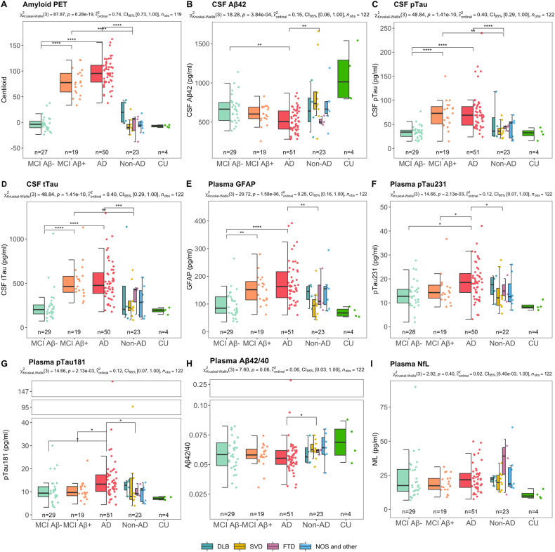
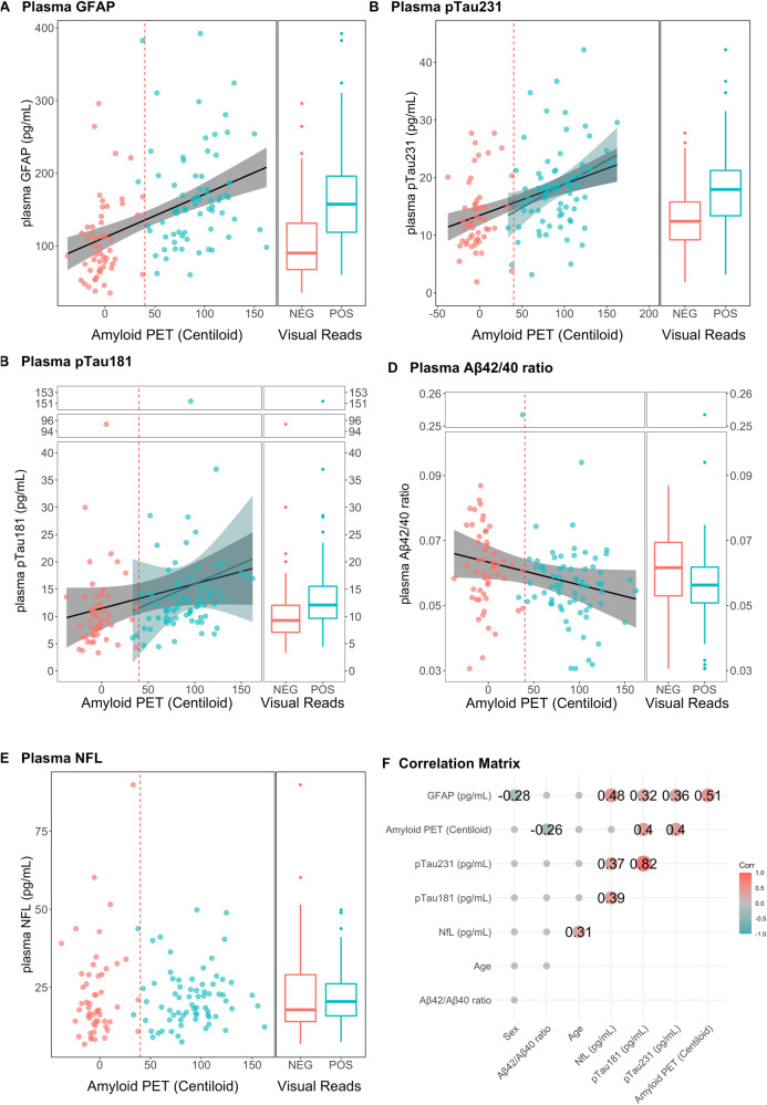
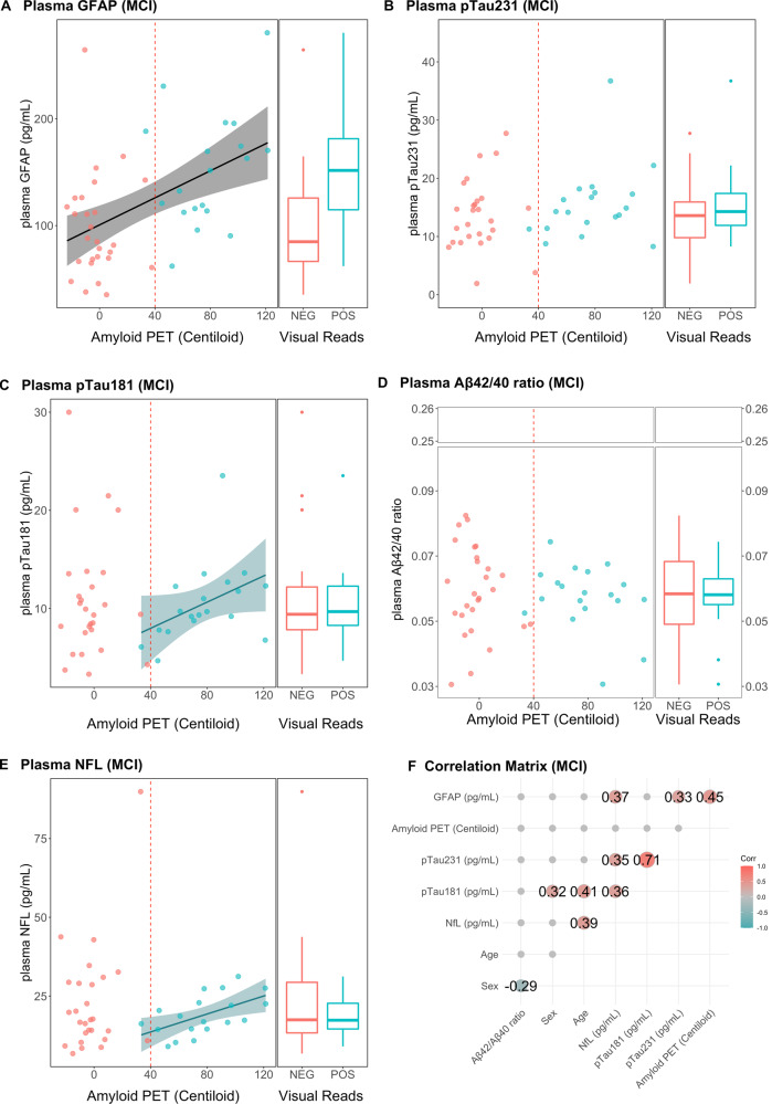
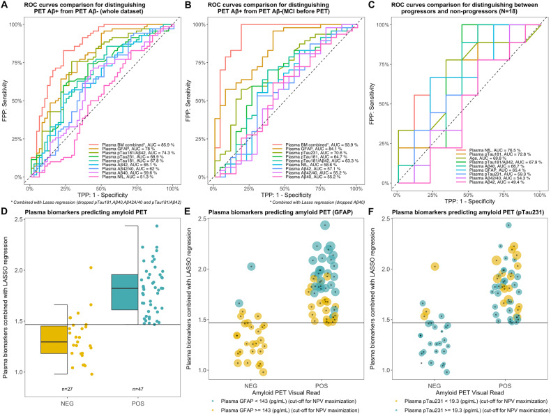
Plasma biomarkers have shown promising performance in research cohorts in discriminating between different stages of Alzheimer's disease (AD). Studies in clinical populations are necessary to provide insights on the clinical utility of plasma biomarkers before their implementation in real-world settings. Here we investigated plasma biomarkers (glial fibrillary acidic protein (GFAP), tau phosphorylated at 181 and 231 (pTau181, pTau231), amyloid β (Aβ) 42/40 ratio, neurofilament light) in 126 patients (age = 65 ± 8) who were admitted to the Clinic for Cognitive Disorders, at Karolinska University Hospital. After extensive clinical assessment (including CSF analysis), patients were classified as: mild cognitive impairment (MCI) (n = 75), AD (n = 25), non-AD dementia (n = 16), no dementia (n = 9). To refine the diagnosis, patients were examined with [18F]flutemetamol PET (Aβ-PET). Aβ-PET images were visually rated for positivity/negativity and quantified in Centiloid. Accordingly, 68 Aβ+ and 54 Aβ- patients were identified. Plasma biomarkers were measured using single molecule arrays (SIMOA). Receiver-operated curve (ROC) analyses were performed to detect Aβ-PET+ using the different biomarkers. In the whole cohort, the Aβ-PET centiloid values correlated positively with plasma GFAP, pTau231, pTau181, and negatively with Aβ42/40 ratio. While in the whole MCI group, only GFAP was associated with Aβ PET centiloid. In ROC analyses, among the standalone biomarkers, GFAP showed the highest area under the curve discriminating Aβ+ and Aβ- compared to other plasma biomarkers. The combination of plasma biomarkers via regression was the most predictive of Aβ-PET, especially in the MCI group (prior to PET, n = 75) (sensitivity = 100%, specificity = 82%, negative predictive value = 100%). In our cohort of memory clinic patients (mainly MCI), the combination of plasma biomarkers was sensitive in ruling out Aβ-PET negative individuals, thus suggesting a potential role as rule-out tool in clinical practice.


Conflict of interest statement

AN has received consulting fee from AVVA Pharmaceuticals, H Lundbeck A/S, Hoffman La Roche, honorarium for lecture Hoffman La Roche, Roche and hold a patent WO 2022/255915. Patent No. PCT/SE2022/050413 PET Tracers. HZ has served at scientific advisory boards and/or as a consultant for Abbvie, Acumen, Alector, Alzinova, ALZPath, Annexon, Apellis, Artery Therapeutics, AZTherapies, CogRx, Denali, Eisai, Nervgen, Novo Nordisk, Optoceutics, Passage Bio, Pinteon Therapeutics, Prothena, Red Abbey Labs, reMYND, Roche, Samumed, Siemens Healthineers, Triplet Therapeutics, and Wave, has given lectures in symposia sponsored by Cellectricon, Fujirebio, Alzecure, Biogen, and Roche, and is a co-founder of Brain Biomarker Solutions in Gothenburg AB (BBS), which is a part of the GU Ventures Incubator Program (outside submitted work). KB has served as a consultant and at advisory boards for Acumen, ALZPath, BioArctic, Biogen, Eisai, Julius Clinical, Lilly, Novartis, Ono Pharma, Prothena, Roche Diagnostics, and Siemens Healthineers; has served at data monitoring committees for Julius Clinical and Novartis; has given lectures, produced educational materials and participated in educational programs for Biogen, Eisai and Roche Diagnostics; and is a co-founder of Brain Biomarker Solutions in Gothenburg AB (BBS), which is a part of the GU Ventures Incubator Program, outside the work presented in this paper.

Figures

Fig. 1
Fig. 1. Levels of Aβ PET (in centiloid), and CSF Alzheimer’s disease biomarkers by diagnostic group.
A PET centiloid was significantly higher in patients diagnosed on AD spectrum. B CSF Ab42 was decreased in patients with AD dementia in comparison to the MCI Aβ– and non-AD dementia groups, whereas no significant difference could be found between MCI Aβ– and Aβ+ groups. In contrast, in the given cohort, levels of CSF pTau and tTau (plots C and D, respectively) were significantly elevated in patients diagnosed on the AD spectrum in relation to patients with non-AD-related pathologies (including comparison between MCI Aβ– and Aβ+ individuals). E Plasma GFAP were statistically different between the groups with minimal (non-AD), intermediate (MCI Aβ + ), and high levels (AD) of Aβ pathology. F, G pTau231 and pTau181 were statistically different between MCI Aβ– and AD in relation to non-AD. H, I No statistically significant difference between the diagnostic groups was observed for plasma NfL and Aβ42/40, except for a significant decrease in plasma Aβ42/40 in AD compared non-AD dementia group. The p values in the subtitles indicate the results of analysis of variance with the Kruskal–Wallis test, between groups, and of post-hoc analysis with Dunn’s test and multiple comparisons correction with the false discovery rate. *p < 0.05, **p < 0.01, ***p < 0.001, ****p < 0.0001.
Fig. 2
Fig. 2. Linear Regressions between plasma biomarkers and Aβ PET (in centiloid) in the whole dataset.
A Plasma GFAP was positively associated with Aβ accumulation in the brain in the whole dataset and in the amyloid-positive and amyloid-negative groups, separately. B, C Plasma pTau181 (B) and pTau181 (C) were positively associated with Aβ accumulation in the brain in the whole dataset and in the amyloid-positive groups. D Plasma NfL was not related to Aβ PET in any group. E Plasma Aβ42/40 was negatively associated with Aβ PET in the whole group only. F Regression lines are drawn only if the Spearman’s rho statistic was significant (p < 0.05). Correlation matrix showing the Spearman’s rho coefficients for the associations between the variables considered.
Fig. 3
Fig. 3. Linear Regressions between plasma biomarkers and Aβ PET (in centiloid) in MCI group.
A Plasma GFAP was the only biomarker positively associated with amyloid accumulation in the brain in the MCI group. B Plasma pTau181, (C) pTau231, D plasma NFL and (E) plasma Aβ42/40 were not related with amyloid PET in the MCI group. C pTau231 was the only biomarker positively associated with amyloid accumulation in the brain in the MCI Aβ+ group. F Regression lines are drawn only if the Spearman’s rho statistic was significant (p < 0.05). Correlation matrix showing the Spearman’s rho coefficients for the associations between the variables considered.
Fig. 4
Fig. 4. Plasma biomarkers as predictors for amyloid PET visual read positivity and conversion to AD.
The combination of biomarkers obtained by LASSO regression first, and plasma GFAP as a single biomarker second, resulted in the two largest AUCs for predicting Aβ positivity in the whole dataset (A) and in the prior to PET MCI group (B). C Plasma NfL was the best predictor for conversion to AD in the MCI Aβ+ group. D, E, F Visualisation of the pooled variable obtained via LASSO regression for the prior to PET MCI group that resulted the best AUC (and specificity and negative predictive values of 100%) from panel (B). D No false positives were identified by the pooled variable. E, F Plasma GFAP and pTau231 values are visualised according to point size, and the colour of the balloons is determined by a threshold for maximisation of the negative predictive value (NPV) (having a test with no false negatives); 143 (pg/ml) and 19.3 (pg/ml) for GFAP and pTau231, respectively.

References

    1. Perini G, Rodriguez-Vieitez E, Kadir A, Sala A, Savitcheva I, Nordberg A. Clinical impact of 18F-FDG-PET among memory clinic patients with uncertain diagnosis. Eur J Nucl Med Mol Imaging. 2021;48:612–22. doi: 10.1007/s00259-020-04969-7. - DOI - PMC - PubMed
    1. Leuzy A, Savitcheva I, Chiotis K, Lilja J, Andersen P, Bogdanovic N, et al. Clinical impact of [18F]flutemetamol PET among memory clinic patients with an unclear diagnosis. Eur J Nucl Med Mol Imaging. 2019;46:1276–86. doi: 10.1007/s00259-019-04297-5. - DOI - PMC - PubMed
    1. Sanchez JS, Hanseeuw BJ, Lopera F, Sperling RA, Baena A, Bocanegra Y, et al. Longitudinal amyloid and tau accumulation in autosomal dominant Alzheimer’s disease: findings from the Colombia-Boston (COLBOS) biomarker study. Alzheimers Res Ther. 2021;13:1–14. doi: 10.1186/s13195-020-00765-5. - DOI - PMC - PubMed
    1. Morris JC, Storandt M, McKeel DW, Rubin EH, Price JL, Grant EA, et al. Cerebral amyloid deposition and diffuse plaques in “normal” aging: Evidence for presymptomatic and very mild Alzheimer’s disease. Neurology. 1996;46:707–19. doi: 10.1212/WNL.46.3.707. - DOI - PubMed
    1. Sperling R, Mormino E, Johnson K. The evolution of preclinical Alzheimer’s disease: Implications for prevention trials. Neuron. 2014;84:608–22. doi: 10.1016/j.neuron.2014.10.038. - DOI - PMC - PubMed

Publication types