Clinical Trial
. 2023 Jul 25;14(1):4180.
doi: 10.1038/s41467-023-39679-9.

Safety and anti-tumour activity of the IgE antibody MOv18 in patients with advanced solid tumours expressing folate receptor-alpha: a phase I trial

Affiliations
Clinical Trial

Safety and anti-tumour activity of the IgE antibody MOv18 in patients with advanced solid tumours expressing folate receptor-alpha: a phase I trial

James Spicer et al. Nat Commun. .

Abstract

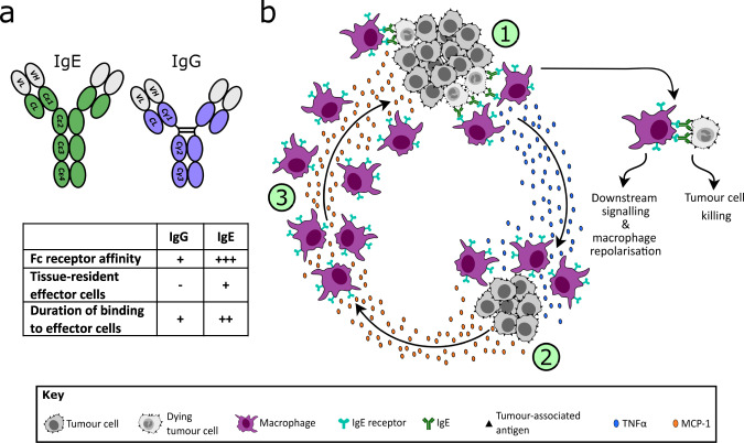
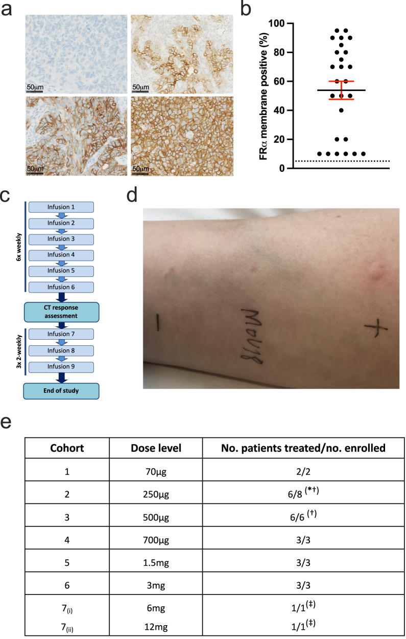
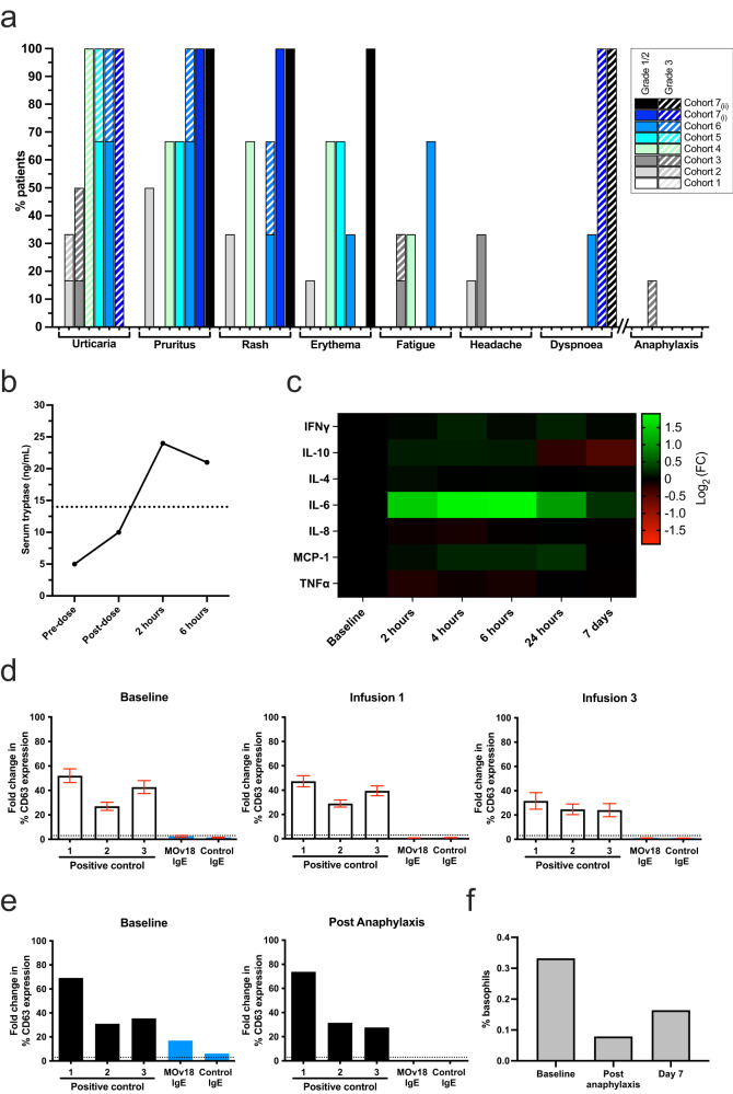
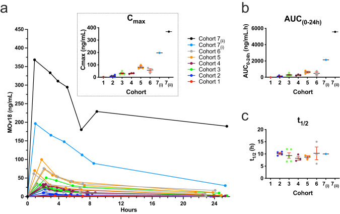
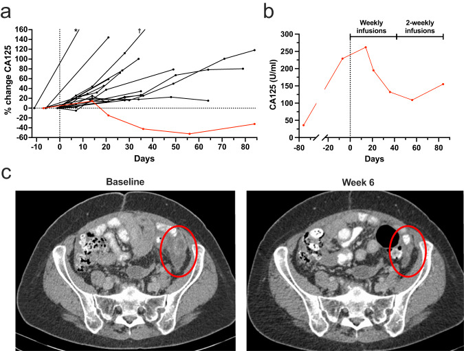
All antibodies approved for cancer therapy are monoclonal IgGs but the biology of IgE, supported by comparative preclinical data, offers the potential for enhanced effector cell potency. Here we report a Phase I dose escalation trial (NCT02546921) with the primary objective of exploring the safety and tolerability of MOv18 IgE, a chimeric first-in-class IgE antibody, in patients with tumours expressing the relevant antigen, folate receptor-alpha. The trial incorporated skin prick and basophil activation tests (BAT) to select patients at lowest risk of allergic toxicity. Secondary objectives were exploration of anti-tumour activity, recommended Phase II dose, and pharmacokinetics. Dose escalation ranged from 70 μg-12 mg. The most common toxicity of MOv18 IgE is transient urticaria. A single patient experienced anaphylaxis, likely explained by detection of circulating basophils at baseline that could be activated by MOv18 IgE. The BAT assay was used to avoid enrolling further patients with reactive basophils. The safety profile is tolerable and maximum tolerated dose has not been reached, with evidence of anti-tumour activity observed in a patient with ovarian cancer. These results demonstrate the potential of IgE therapy for cancer.


Conflict of interest statement

J.S. and S.K. are co-founders of Epsilogen Ltd. H.B. is presently employed, and J.C. formerly employed, through a fund from Epsilogen Ltd. S.K., H.B., H.G., D.J, G.P. and J.S. hold patents on anti-tumour IgE antibodies. The remaining authors declare no competing interests.

Figures

Fig. 1
Fig. 1. Mechanism of cytotoxicity mediated by MOv18 IgE.
a The structurally distinct Fc domains of IgE and IgG underpin differences in their biological characteristics, supporting exploration of IgE therapies as potentially superior to IgG [refs. ,]. b (1) IgE bound to Fc receptors on macrophages is crosslinked by antigen expressed on tumour cells, leading to target cell cytotoxicity, release of proinflammatory mediators (e.g. TNFα) and macrophage repolarization. (2) TNFα upregulation in turn triggers MCP-1 production by monocytes and tumour cells, followed by (3) recruitment of further macrophages, mediated by MCP-1 [refs. ,,].
Fig. 2
Fig. 2. Patient selection and study treatment.
a Representative immunohistochemistry of paraffin-embedded ovarian cancers showing a range of FRα membrane expression on 10 to 95% of tumour cells. b FRα membrane expression for all enroled patients (mean ± SEM; dotted line represents the 5% expression threshold required for trial eligibility; n = 26). c The trial dosing schedule consisted of 6 weekly doses of MOv18 IgE, before first on-treatment tumour assessment. d Skin prick testing comprised histamine positive control [+], saline negative control [−] and MOv18 IgE solution. The result of this typical skin prick test was negative. e MOv18 IgE dose escalation, and number of patients treated in each cohort (*cohort 2 included 2 patients not evaluable for safety who did not receive intravenous dosing because of positive intradermal tests (see text); a DLT occurred in these cohorts; intra-patient dose escalation). Source data are provided as a Source Data file.
Fig. 3
Fig. 3. Treatment-related adverse events, and basophil activation test (BAT) as a predictor of anaphylaxis.
a Adverse events (AEs) experienced by ≥10% of patients (as well as anaphylaxis in 1/26 (4%)) are grouped according to severity and by dose cohort (1–7(ii) from left to right for each AE). No related AEs occurred at the lowest dose (cohort 1), and there was no grade 4 or 5 toxicity at any dose. Acute hypotension responding promptly to intramuscular adrenaline was seen in 1 patient treated at 500 μg (cohort 3). b Serum tryptase was elevated in this patient at 2 and 6 h following the infusion, supporting a diagnosis of drug-related anaphylaxis to explain this adverse event (dotted line represents upper limit of normal for serum tryptase [14 ng/ml]). c Fold change (Log2) in median circulating cytokine levels post-dose (2, 6, 24 h and 7 days) relative to normalised baseline values for each patient (n = 26). d Marked upregulation of the basophil activation marker CD63 in response to ex vivo stimulation of patient blood using positive controls (1 = anti-FcεRI, 2 = fMLP, 3 = anti-IgE), but not to MOv18 or control IgEs. This BAT result (n = 26) remained negative when performed following the 1st (n = 24) and 3rd (n = 20) MOv18 IgE doses (mean ± SEM) for all trial subjects excluding the single patient who experienced anaphylaxis. Negative control IgE was not included in all assays. e By contrast, the baseline BAT was positive for the patient experiencing anaphylaxis at the time of their subsequent MOv18 IgE infusion (n = 1). Unlike all other patients, MOv18 IgE antibody produced a 17-fold increase in basophil activation in this individual’s blood at baseline (left panel, blue bars). The BAT for this patient became negative after the episode of anaphylaxis (right panel), likely as a result of basophil depletion. f Peripheral basophil counts fell immediately following MOv18 IgE infusion and anaphylaxis in this patient, but not in other trial subjects after their MOv18 IgE dosing (Supplementary Fig. 1), recovering by day 7 (n = 1). Source data are provided as a Source Data file.
Fig. 4
Fig. 4. Plasma pharmacokinetics of MOv18 IgE.
a Linear plot of plasma drug concentrations for evaluable patients (n = 23) showing dose-proportional Cmax (inset; mean ± SEM; n = 23), which exceeded baseline endogenous IgE levels in 25% of patients. b Area under the curve (AUC(0–24h); mean ± SEM; n = 20) also increased with dose. c Half-life (mean ± SEM; n = 19) was consistent in the range of doses tested, with a mean t1/2 = 9.4 h across all cohorts. Some measurements were below level of quantification for MOv18 IgE. Source data are provided as a Source Data file.
Fig. 5
Fig. 5. Anti-tumour activity.
a Percentage change in CA125 circulating tumour marker for evaluable patients (n = 17; vertical dotted line indicates initiation of dosing; red line indicates a patient with marked CA125 reduction and tumour volume reduction; * and  = tumour marker progression >200%). b Marked but short-lived CA125 reduction following MOv18 IgE dosing in a patient with progression prior to treatment. CA125 relapse coincided with change from weekly to alternate-weekly dosing after 6 weeks. c Pelvic CT images for this patient at baseline and week 6 of treatment showing reduction of peritoneal tumour deposit (red oval). Source data are provided as a Source Data file.

References

    1. Kretschmer A, Schwanbeck R, Valerius T, Rosner T. Antibody isotypes for tumor immunotherapy. Transfus. Med. Hemother. 2017;44:320–326. doi: 10.1159/000479240. - DOI - PMC - PubMed
    1. Mukai K, Tsai M, Starkl P, Marichal T, Galli SJ. IgE and mast cells in host defense against parasites and venoms. Semin. Immunopathol. 2016;38:581–603. doi: 10.1007/s00281-016-0565-1. - DOI - PMC - PubMed
    1. Sutton, B.J., Davies, A.M., Bax, H.J. & Karagiannis, S.N. IgE antibodies: from structure to function and clinical translation. Antibodies (Basel)8 (2019). - PMC - PubMed
    1. Hagan P, Blumenthal UJ, Dunn D, Simpson AJ, Wilkins HA. Human IgE, IgG4 and resistance to reinfection with Schistosoma haematobium. Nature. 1991;349:243–245. doi: 10.1038/349243a0. - DOI - PubMed
    1. Hsiao HC, Fan X, Jordan RE, Zhang N, An Z. Proteolytic single hinge cleavage of pertuzumab impairs its Fc effector function and antitumor activity in vitro and in vivo. Breast Cancer Res. 2018;20:43. doi: 10.1186/s13058-018-0972-4. - DOI - PMC - PubMed

Publication types

Associated data