. 2023 Oct;165(10):2873-2883.
doi: 10.1007/s00701-023-05687-3. Epub 2023 Jul 26.

Metastatic meningioma: a case series and systematic review

Affiliations

Metastatic meningioma: a case series and systematic review

Vratko Himič et al. Acta Neurochir (Wien). 2023 Oct.

Abstract

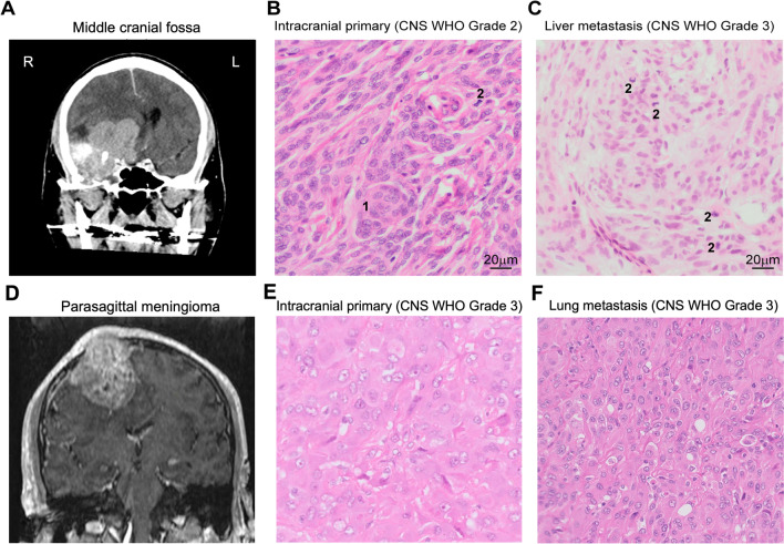
Background: Meningiomas are the most common primary intracranial tumor. While the majority of meningiomas are benign, rarely they can metastasize extracranially. There is a need for a more comprehensive review of these patients to improve our understanding of this rare phenomenon and its prevalence globally. Here we describe our institution's experience of patients presenting with metastatic meningiomas. We further perform a systematic review of the existing literature to explore common features of this rare manifestation of meningioma and review the efficacy of current treatments.

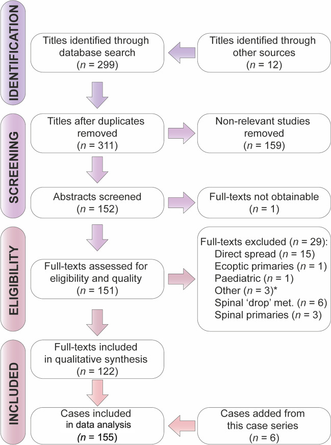
Methods: We performed a retrospective clinical review of all adult patients with metastatic meningioma managed at our institution over the past 20 years, identifying 6 patients. We then performed a systematic review of cases of metastatic meningioma in the literature ranging from the years 1886 to 2022. A descriptive analysis was then conducted on the available data from 1979 onward, focusing on the grade and location of the primary tumor as well as the latency period to, and location of, the metastasis.

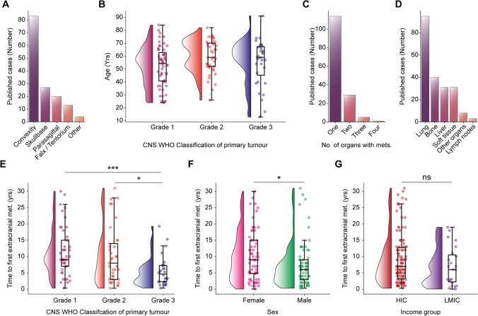
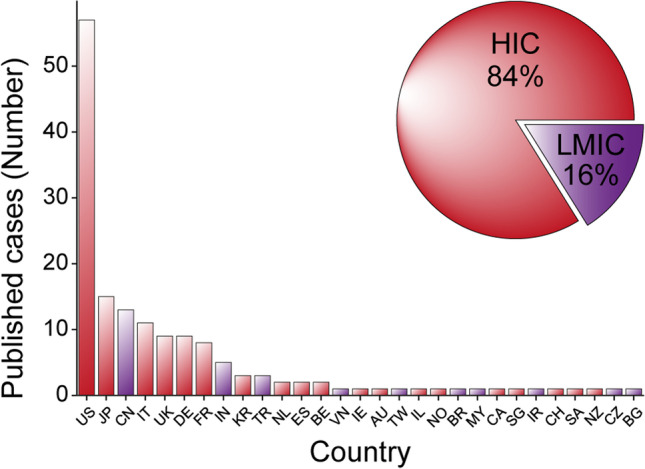
Results: In total, we analyzed 155 cases. Fifty-four percent of patients initially presented with a primary meningioma located in the convexity. The most common site of metastasis was the lung. Risk factors associated with a shorter time to metastasis were male sex and a high initial grade of the tumor. Regarding treatment, the addition of chemotherapy was the most common adjunct to the standard management of surgery and radiotherapy. Despite an exhaustive review we were unable to identify effective treatments. The majority of published cases came from centers situated in high-income countries (84%) while only 16% came from lower- and middle-income countries.

Conclusions: Metastatic meningiomas pose a pertinent, and likely underestimated, clinical challenge within modern neurosurgery. To optimize management, timely identification of these patients is important. More research is needed to explore the mechanisms underlying these tumors to better guide the development of effective screening and management protocols. However, screening of each meningioma patient is not feasible, and at the heart of this challenge is the inability to control the primary disease. Ultimately, a consensus is needed as to how to correctly screen for and manage these patients; genomic and epigenomic approaches could hold the answer to finding druggable targets.

Keywords: (Epi)genomics; Meningioma; Metastatic; Oncology; Palliative; Recurrent; Screening.


Conflict of interest statement

The authors declare co competing interests.

Figures

Fig. 1
Fig. 1
Two example cases. A–C Extracranial metastasis to the liver. A A CT scan with gadolinium showing an intracranial primary meningioma before surgery at the right middle cranial fossa (case 5). B Photomicrograph of hematoxylin and eosin (HE)-stained section of temporal resection showing characteristic whorl (1) and a single mitotic figure (2). Mitotic activity was present between four and 20 per 10 high power fields. The primary tumor was diagnosed as an atypical intracranial meningioma and classified as CNS WHO grade 2. C Photomicrograph of HE-stained section of liver biopsy showing four mitotic figure (2). Mitotic activity is present in more than 20 per 10 high power fields. Tumor was diagnosed as a metastatic anaplastic meningioma and classified as CNS WHO grade 3. D–F Extracranial metastasis to the lung. D A T1-weighted MRI scan showing a parasagittal primary meningioma with demonstration of the patient’s presenting complaint; a skin “lump” due to the extension of the neoplasm (case 3). E and F Both the primary (E) and lung metastasis (F) show an anaplastic meningioma, CNS WHO grade 3 (mitotic activity > 20 per 10 high power fields). Sheet-like architecture and some rhabdoid morphology can be seen
Fig. 2
Fig. 2
PRISMA flowchart. “Other” (*) includes patients who were initially classified as having primary intracranial meningiomas which were later found to be hemangiopericytomas on histology
Fig. 3
Fig. 3
Age distribution, grade, and location of primary intracranial tumor. A Location of primary intracranial meningioma. B No difference in age of patients presenting with different CNS WHO grades of primary intracranial meningioma (grade 1: mean: 54.5 ± SEM 2.1 years; grade 2: mean: 59.59 ± SEM: 2.0 years; grade 3: mean 55.1 ± SEM 3.1 years; p = 0.57, one-way ANOVA test). C Bar plot of number of cases which presented with a single organ or multiple organs containing extracranial metastases. D The number of cases in which metastases were found in specific organs. E The CNS WHO grade of the primary intracranial meningioma significantly affects the time to first presentation with an extracranial metastasis (p = 0.0003, Kruskal–Wallis test). Patients with CNS WHO grade 3 primary intracranial meningiomas present with extracranial metastases sooner compared to those with CNS WHO grade 1 (grade 3: median 4.4 IQR 5.0 years vs grade 1: median 9 IQR 8.5 years, p = 0.0002, Dunn’s multiple comparisons test) or CNS WHO grade 2 (grade 2: median 8 IQR 11.5 years, p = 0.02, Dunn’s multiple comparisons test). There was no difference between grade 1 and grade 2 tumors (p = 0.76, Dunn’s multiple comparisons test). F Differences in sex and time to first extracranial metastatic lesion (female: median 9.0 IQR 10.3 years vs male: median 6.0 IQR 8.75 years, p = 0.0003, Mann–Whitney test). G Differences time to first extracranial metastatic lesion and the income group of the country in which the patient was treated (high-come country, HIC: median 7.0 IQR 10.0 years vs low-middle-income country, LMIC: median 6 IQR 8 years, p = 0.16 Mann–Whitney test)
Fig. 4
Fig. 4
Geographic and economic distribution of published cases. The bar plot stratifies the number of cases published from each country. The pie plot (inset) separates cases as per the income group of the country from which the institution they came from is situated. Income groups where defined as per data from the World Bank based on gross national income (GNI) per capita countries were either classified as high-income (HIC) or low-middle-income (LMIC) which includes low-income, lower-middle income, and upper-middle-income

Similar articles

Cited by

References

    1. Alexandru D, Glantz MJ, Kim L, Chamberlain MC, Bota DA. Pulmonary metastases in patients with recurrent, treatment-resistant meningioma: prognosis and identification by 111Indium-octreotide imaging. Cancer. 2011;117(19):4506–4511. doi: 10.1002/cncr.26065. - DOI - PubMed
    1. Bayley JC, Hadley CC, Harmanci AO, Harmanci AS, Klisch TJ, Patel AJ. Multiple approaches converge on three biological subtypes of meningioma and extract new insights from published studies. Sci Adv. 2022;8(5):eabm6247. doi: 10.1126/sciadv.abm6247. - DOI - PMC - PubMed
    1. Bi WL, Greenwald NF, Abedalthagafi M, et al. Genomic landscape of high-grade meningiomas. Npj Genomic Med. 2017;2(1):1–14. doi: 10.1038/s41525-017-0014-7. - DOI - PMC - PubMed
    1. Brodbelt AR, Barclay ME, Greenberg D, Williams M, Jenkinson MD, Karabatsou K. The outcome of patients with surgically treated meningioma in England: 1999–2013. A cancer registry data analysis. Br J Neurosurg. 2019;33(6):641–647. doi: 10.1080/02688697.2019.1661965. - DOI - PubMed
    1. Chew SJY, Rajesvaran C, Woo X, Goh CH. Atypical meningioma with nodal metastasis: a case report. Malays J Pathol. 2021;43(3):453–456. - PubMed

Publication types