. 2024 Feb;49(3):497-507.
doi: 10.1038/s41386-023-01659-4. Epub 2023 Jul 25.

Puerarin attenuates valproate-induced features of ASD in male mice via regulating Slc7a11-dependent ferroptosis

Affiliations

Puerarin attenuates valproate-induced features of ASD in male mice via regulating Slc7a11-dependent ferroptosis

Peiyan Jiang et al. Neuropsychopharmacology. 2024 Feb.

Abstract

Autism spectrum disorder (ASD) is a complicated, neurodevelopmental disorder characterized by social deficits and stereotyped behaviors. Accumulating evidence suggests that ferroptosis is involved in the development of ASD, but the underlying mechanism remains elusive. Puerarin has an anti-ferroptosis function. Here, we found that the administration of puerarin from P12 to P15 ameliorated the autism-associated behaviors in the VPA-exposed male mouse model of autism by inhibiting ferroptosis in neural stem cells of the hippocampus. We highlight the role of ferroptosis in the hippocampus neurogenesis and confirm that puerarin treatment inhibited iron overload, lipid peroxidation accumulation, and mitochondrial dysfunction, as well as enhanced the expression of ferroptosis inhibitory proteins, including Nrf2, GPX4, Slc7a11, and FTH1 in the hippocampus of VPA mouse model of autism. In addition, we confirmed that inhibition of xCT/Slc7a11-mediated ferroptosis occurring in the hippocampus is closely related to puerarin-exerted therapeutic effects. In conclusion, our study suggests that puerarin targets core symptoms and hippocampal neurogenesis reduction through ferroptosis inhibition, which might be a potential drug for autism intervention.


Conflict of interest statement

The authors declare no competing interests.

Figures
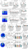
Fig. 1
Fig. 1. PU could ameliorate VPA-induced autistic-like behaviors and cognitive impairment in male mice.
A A 7-day body weight record. B Self-grooming time of experimental mice. C The number of buried marbles. D Pattern diagram of social approach test. Warmer colors (red) indicate plenty of time spent exploring by the experimental mice. E Statistical graph of time spent sniffing the novel mice or object. F The preference index (S1-O) from sniffing time. G Pattern diagram of social recognition test. H Statistical graph of time spent sniffing the novel mice or the familiar mice. I The preference index (S2–S1) from sniffing time. J Nest score in nesting test. K Discrimination index of novel object recognition. L, M A correct number of spontaneous rotations in the Y-maze test. N Pattern diagram of MWM. O Escape latency during 5 days of training. P Time spent in the target or opposite quadrant. Q The number of crossing the target quadrant. R Pattern diagram of reverse MWM. S Escape latency during 3 days of training. T Time spent in the target or opposite quadrant. U The number of crossing the target quadrant. Statistical method: repeated-measures ANOVA: A, O, S; one-way ANOVA : B, C, F, I; paired t-test: E, H, P, T; two-way ANOVA: J, K, L, M, Q, U P < 0.05, **P < 0.01, and ***P < 0.001. In (A,O,S) # represents Sal + DMSO vs VPA + DMSO; #P < 0.05, ##P < 0.01, and ###P < 0.001, *represent VPA + DMSO and VPA + PU. *P < 0.05, **P < 0.01, and ***P < 0.001.
Fig. 2
Fig. 2. PU treatment improves decreased neurogenesis caused by P14VPA treatment in the DG of the P15 hippocampus.
A The Sox2+ cells in the DG of the P15 hippocampus were presented in the first row. The next row exhibited GFAP+ cells in the DG. Furthermore, the third row showed representative images of Sox2 (red)-and GFAP (green)-double-positive cells in the DG. Scale bar: 100 μm; magnificent figure: 20 μm. B Representative images of DCX. Scale bar:20 μm. C Protein expressions of GFAP and DCX in the P15 hippocampus were detected by WB. D Statistical graph of the SOX2 and GFAP double-positive cells in the DG. E Statistical graph of the SOX2+ cells in the DG. F Statistical of GFAP protein expression level. G Statistical of DCX protein expression level. Statistical method: two-way ANOVA: DG. *P < 0.05, **P < 0.01, and ***P < 0.001.
Fig. 3
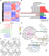
Fig. 3. The target xCT/Slc7a11 can be a bridge connecting hippocampus neurogenesis and ferroptosis in the effect of PU.
A Hierarchical clustering heat map of the four groups. B KEGG enrichment of the 5203 genes co-regulated by VPA and PU. C GO enrichment of the 5203 genes co-regulated by VPA and PU. D Cluster analysis of related biological processes. E Verification of high variation genes in RNA-seq by RT-PCR. Statistical method:two-way ANOVA: E *P < 0.05, **P < 0.01, and ***P < 0.001.
Fig. 4
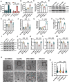
Fig. 4. Characteristics of ferroptosis in the P15 hippocampus of VPA-treated male mice were ameliorated by PU treatment.
A Content of ferrous and total iron in the P15 hippocampus. B The MDA level in the P15 hippocampus. C The glutathione concentration in the P15 hippocampus. D Protein expression of 4-HNE and ACSL4 detected by Western blot. E, F Statistical graphs show the 4-HNE and ACSL4 protein expression levels. G, H Nrf2 and Slc7a11 mRNA levels were detected by RT-PCR. I Protein expressions of Nrf2, Slc7a11, FTH1, and GPX4 in the P15 hippocampus. JM Statistical graphs show the protein expression level. N Electron microscope of mitochondrial morphology in the P15 hippocampus. The second row represents a partial enlargement of the first row, respectively. The black arrow shows crumpled mitochondria. O Statistical graph of mitochondrial area. (n = 3 mice per group, 100 mitochondria per mouse) Statistical method: two-way ANOVA: AO. *P < 0.05, **P < 0.01, and ***P < 0.001.
Fig. 5
Fig. 5. Erastin blocked the improved effect of PU in VPA-induced ASD phenotypes.
A Ferroptosis-related proteins (GPX4, FTH1, and Slc7a11) levels in the P15 hippocampus. BD The protein levels of GPX4, FTH1, and Slc7a11 in the P15 hippocampus. E Glutathione level in the P15 hippocampus. F SOX2 protein levels in the P15 hippocampus of four groups were detected by WB. G Statistical graph of the SOX2 protein level. H Time spent in self-grooming. I Statistic of the number of buried marbles. J Preference Index (S1-O) from sniffing time in the social approach phase. K Sniffing time on novel mouse or object. L Preference Index (S2–S1) from sniffing time in the social recognition phase. M Sniffing time on novel mouse or familiar mouse. N Morphology of neural stem cell spheres in primary culture and their identification staining with SOX2. O C11 BODIPY staining detected by Flow Cytometry. P Statistical results of C11 BODIPY staining. Q Statistical results of CCK-8. R C11 BODIPY staining detected by Confocal Microscope. Statistical method: two-way ANOVA: BE, G; paired t-test: K, M; one-way ANOVA: HJ, L, P, Q. *P < 0.05, **P < 0.01, and ***P < 0.001.

References

    1. Lord C, Charman T, Havdahl A, Carbone P, Anagnostou E, Boyd B, et al. The Lancet Commission on the future of care and clinical research in autism. Lancet (Lond, Engl) 2022;399:271–334. doi: 10.1016/S0140-6736(21)01541-5. - DOI - PubMed
    1. Newschaffer CJ, Croen LA, Daniels J, Giarelli E, Grether JK, Levy SE, et al. The epidemiology of autism spectrum disorders. Annu Rev Public Health. 2007;28:235–58. doi: 10.1146/annurev.publhealth.28.021406.144007. - DOI - PubMed
    1. Nestler EJ, Hyman SE. Animal models of neuropsychiatric disorders. Nat Neurosci. 2010;13:1161–9. doi: 10.1038/nn.2647. - DOI - PMC - PubMed
    1. Sharma SR, Gonda X, Tarazi FI. Autism Spectrum Disorder: Classification, diagnosis and therapy. Pharmacol Ther. 2018;190:91–104. doi: 10.1016/j.pharmthera.2018.05.007. - DOI - PubMed
    1. Liu G, Yu Q, Tan B, Ke X, Zhang C, Li H, et al. Gut dysbiosis impairs hippocampal plasticity and behaviors by remodeling serum metabolome. Gut Microbes. 2022;14:2104089. doi: 10.1080/19490976.2022.2104089. - DOI - PMC - PubMed