

Self-expandable metallic stent-induced esophagorespiratory fistulas in patients with advanced esophageal cancer
- PMID: 37491991
- PMCID: PMC10665617
- DOI: 10.5946/ce.2022.297
Self-expandable metallic stent-induced esophagorespiratory fistulas in patients with advanced esophageal cancer
Abstract
Background/aims: Self-expandable metallic stents (SEMSs) are widely adopted for the palliation of dysphagia in patients with malignant esophageal strictures. An important adverse event is the development of SEMS-induced esophagorespiratory fistulas (SEMS-ERFs). This study aimed to assess the risk factors related to the development of SEMS-ERF after SEMS placement in patients with esophageal cancer.
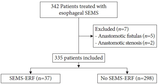
Methods: This retrospective study was performed at the Instituto do Cancer do Estado de São Paulo. All patients with malignant esophageal strictures who underwent esophageal SEMS placement between 2009 and 2019 were included in the study.
Results: Of the 335 patients, 37 (11.0%) developed SEMS-ERF, with a median time of 129 days after SEMS placement. Stent flare of 28 mm (hazard ratio [HR], 2.05; 95% confidence interval [CI], 1.15-5.51; p=0.02) and post-stent chemotherapy (HR, 2.0; 95% CI, 1.01-4.00; p=0.05) were associated with an increased risk of developing SEMS-ERF, while lower-third tumors were a protective factor (HR, 0.5; 95% CI, 0.26-0.85; p=0.01). No difference was observed in overall survival.
Conclusion: The incidence of SEMS-ERFs was 11%, with a median time of 129 days after SEMS placement. Post-stent chemotherapy and a 28 mm stent flare were associated with a higher risk of SEMS-ERF.
Keywords: Esophageal fistula; Esophageal neoplasms; Self-expandable metallic stents.
Conflict of interest statement
The authors have no potential conflicts of interest.
Figures


Comment in
-
How to reduce fistula formation after self-expandable metallic stent insertion for treating malignant esophageal stricture?Clin Endosc. 2023 Nov;56(6):735-737. doi: 10.5946/ce.2023.257. Epub 2023 Nov 17. Clin Endosc. 2023. PMID: 37981742 Free PMC article. No abstract available.
References
-
- Parker RK, White RE, Topazian M, et al. Stents for proximal esophageal cancer: a case-control study. Gastrointest Endosc. 2011;73:1098–1105. - PubMed
-
- Medeiros VS, Martins BC, Lenz L, et al. Adverse events of self-expandable esophageal metallic stents in patients with long-term survival from advanced malignant disease. Gastrointest Endosc. 2017;86:299–306. - PubMed
-
- Homann N, Noftz MR, Klingenberg-Noftz RD, et al. Delayed complications after placement of self-expanding stents in malignant esophageal obstruction: treatment strategies and survival rate. Dig Dis Sci. 2008;53:334–340. - PubMed
-
- Lenz CJ, Bick BL, Katzka D, et al. Esophagorespiratory fistulas: survival and outcomes of treatment. J Clin Gastroenterol. 2018;52:131–136. - PubMed
LinkOut - more resources
Full Text Sources
