. 2023 Aug 3;127(30):6597-6607.
doi: 10.1021/acs.jpcb.3c00976. Epub 2023 Jul 26.

Self-Assembly of Insulin-Derived Chimeric Peptides into Two-Component Amyloid Fibrils: The Role of Coulombic Interactions

Affiliations

Self-Assembly of Insulin-Derived Chimeric Peptides into Two-Component Amyloid Fibrils: The Role of Coulombic Interactions

Mateusz Fortunka et al. J Phys Chem B. .

Abstract

Canonical amyloid fibrils are composed of covalently identical polypeptide chains. Here, we employ kinetic assays, atomic force microscopy, infrared spectroscopy, circular dichroism, and molecular dynamics simulations to study fibrillization patterns of two chimeric peptides, ACC1-13E8 and ACC1-13K8, in which a potent amyloidogenic stretch derived from the N-terminal segment of the insulin A-chain (ACC1-13) is coupled to octaglutamate or octalysine segments, respectively. While large electric charges prevent aggregation of either peptide at neutral pH, stoichiometric mixing of ACC1-13E8 and ACC1-13K8 triggers rapid self-assembly of two-component fibrils driven by favorable Coulombic interactions. The low-symmetry nonpolar ACC1-13 pilot sequence is crucial in enforcing the fibrillar structure consisting of parallel β-sheets as the self-assembly of free poly-E and poly-K chains under similar conditions results in amorphous antiparallel β-sheets. Interestingly, ACC1-13E8 forms highly ordered fibrils also when paired with nonpolypeptide polycationic amines such as branched polyethylenimine, instead of ACC1-13K8. Such synthetic polycations are more effective in triggering the fibrillization of ACC1-13E8 than poly-K (or poly-E in the case of ACC1-13K8). The high conformational flexibility of these polyamines makes up for the apparent mismatch in periodicity of charged groups. The results are discussed in the context of mechanisms of heterogeneous disease-related amyloidogenesis.


Conflict of interest statement

The authors declare no competing financial interest.

Figures

Figure 1
Figure 1
(A) Design of the ACC1–13E8 and ACC1–13K8 peptides. The amino acid sequence of the N-terminal segment of bovine insulin’s A-chain (the first 13 residues) was extended at the C-end by additional 8 glutamate or 8 lysine residues. (B) Nonbranched and branched structures of monomer units of PAA and PEI, respectively. Averaged degrees of polymerization are given in parentheses.
Figure 2
Figure 2
ThT fluorescence-based screening for amyloidal aggregates formed upon the mixing of aqueous solutions of selected oligocations and oligoanions at pH 7 and subsequent incubation. Numerical values superimposed on the UV-illuminated plate image correspond to ThT emission readouts (λex. 440 nm/λem. 485 nm) collected for wells filled with nonmixed (top row and far left column) and mixed (within the blue frame) solutions of specified compounds after 48 h of incubation at 37 °C. The final concentration of ACC1–13E8 was 0.5 mg/mL, while the concentrations of added counterions were calculated assuming a 1:1 charge compensation stoichiometry and full ionization of all carboxyl and amine (primary and secondary) groups. Each well contained ThT at a 30 μM concentration. The most fluorescing samples are indicated with green rings; the control readout for the neat ThT solution is marked with a red ring.
Figure 3
Figure 3
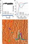
Cofibrillization of ACC1–13E8 and ACC1–13K8. (A) ThT fluorescence-based monitoring of fibrillization of 0.22 mM aqueous solutions of ACC1–13E8, ACC1–13K8, and their equimolar mixture; pH 7, 20 μM ThT, 37 °C, 300 rpm, 24 h. (B) Far-UV CD spectra of aqueous suspension of ACC1–13E8-ACC1–13K8 coaggregate juxtaposed with the spectra of individual peptides at pH 7 (0.11 mM conc., 1 mm optical pathway). (C) Amplitude AFM image of ACC1–13E8-ACC1–13K8 coaggregate; overlaid are cross sections of the selected fibrillar specimen.
Figure 4
Figure 4
Time-lapse ATR FT-IR spectra (amide I band region) of an equimolar mixture of ACC1–13E8 and ACC1–13K8, pH 7, undergoing spontaneous coaggregation while incubated in a thermoblock at 37 °C.
Figure 5
Figure 5
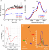
Coaggregation of ACC1–13E8 and PAA. (A) ThT fluorescence trajectories obtained for samples containing ACC1–13E8 at fixed concentrations of 0.22 mM and various concentrations of PAA, as indicated. Other conditions of fibrillization: pH 7, 30 μM ThT, 37 °C, 300 rpm, 24 h. (B) FT-IR spectra of dry coaggregates collected afterward. (C) Far-UV CD spectra of aqueous suspension of ACC1–13E8-PAA coaggregate and neat ACC1–13E8 both at pH 7 (0.11 peptide mM conc., 1 mm optical pathway). (D) Amplitude AFM image of ACC1–13E8-PAA coaggregates; overlaid are cross sections of the selected fibrillar specimen.
Figure 6
Figure 6
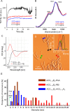
Coaggregation of ACC1–13E8 and PEI; comparison of the thickness of various aggregates. (A) ThT fluorescence trajectories obtained for samples containing fixed concentrations of ACC1–13E8 (0.22 mM) and various indicated concentrations of PEI. Other conditions of fibrillization: pH 7, 30 μM ThT, 37 °C, 300 rpm, 24 h. (B) FT-IR spectra of dry coaggregates collected afterward. (C) Far-UV CD spectra of aqueous suspension of ACC1–13E8-PEI coaggregate and neat ACC1–13E8 both at pH 7 (0.11 peptide mM conc., 1 mm optical pathway). (D) Amplitude AFM image of ACC1–13E8-PEI coaggregate; overlaid are cross sections of the selected fibrillar specimen. (E) Histogram representation of relative abundancies of fibrillar specimens of various widths (estimated according to the AFM height images) in aggregates of ACC1–13E8-ACC1–13K8, ACC1–13E8-PAA, and ACC1–13E8-PEI.
Figure 7
Figure 7
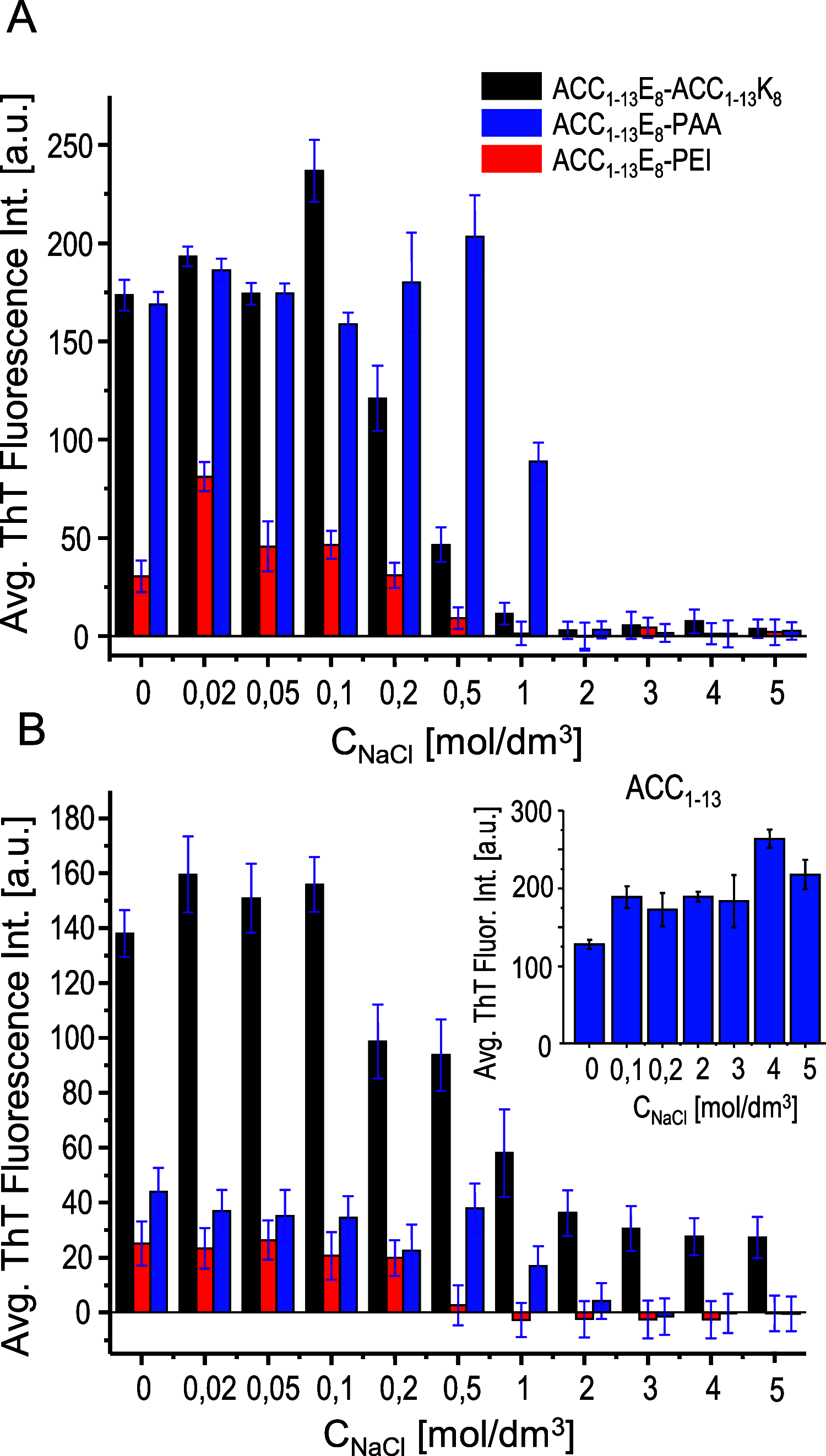
Influence of ionic strength on ACC1–13E8-polycation cofibrillization and on stability of preformed coaggregates. (A) ThT emission readouts (λex. 440 nm/λem. 485 nm, 30 μM ThT) averaged over the third hour of incubation at 37 °C of ACC1–13E8 (0.5 mg/mL) mixed with ACC1–13K8, PAA, or PEI in the presence of increasing NaCl concentration. The mixing stoichiometry of all negatively and positively ionized groups was 1:1, assuming full ionization of all carboxyl and amine (primary and secondary) groups (the same conditions as in Figure 2), pH 7. (B) ThT emission readouts of ACC1–13E8-polycation coaggregates formed under typical conditions (1:1 ionic stoichiometry of mixing, pH 7, 24 h incubation at 37 °C, without NaCl) and subsequently transferred to aqueous NaCl solutions of specified concentrations, pH 7, containing 30 μM ThT. The data correspond to emission values averaged over 18 h of incubation at 37 °C. The control data on the NaCl effect on ThT-stained fibrils of ACC1–13 incubated under analogous conditions is shown in the inset.
Figure 8
Figure 8
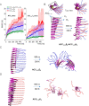
MD-based analysis of stability of ACC1–13E8-ACC1–13K8 coaggregate. (A) Time-dependent changes of RMSD of Cα atoms at 300 K in preassembled fibrillar stacks of extended peptides chains (in-register parallel β-sheet structure): alternate layers of ACC1–13E8 and ACC1–13K8 versus analogous structures composed of ACC1–13E8 and ACC1–13K8 only. The presented averaged RMSD trajectories have been calculated for all Cα atoms (left) and Cα atoms within the ACC1–13 segments only (right). (B–D) Snapshots of the initial and final (after 500 ns) states of these structures.

Similar articles

Cited by

References

    1. Ke P. C.; Zhou R.; Serpell L. C.; Riek R.; Knowles T. P. J.; Lashuel H. A.; Gazit E.; Hamley I. W.; Davis T. P.; Fändrich M.; et al. Half a Century of Amyloids: Past, Present and Future. Chem. Soc. Rev. 2020, 49, 5473–5509. 10.1039/C9CS00199A. - DOI - PMC - PubMed
    1. Fändrich M.; Fletcher M. A.; Dobson C. M. Amyloid Fibrils from Muscle Myoglobin. Nature 2001, 410, 165–166. 10.1038/35065514. - DOI - PubMed
    1. Fandrich M.; Dobson C. M. The Behaviour of Polyamino Acids Reveals an Inverse Side Chain Effect in Amyloid Structure Formation. EMBO J. 2002, 21, 5682–5690. 10.1093/emboj/cdf573. - DOI - PMC - PubMed
    1. Buell A. K. The Growth of Amyloid Fibrils: Rates and Mechanisms. Biochem. J. 2019, 476, 2677–2703. 10.1042/BCJ20160868. - DOI - PubMed
    1. Iadanza M. G.; Jackson M. P.; Hewitt E. W.; Ranson N. A.; Radford S. E. A New Era for Understanding Amyloid Structures and Disease. Nat. Rev. Mol. Cell Biol. 2018, 19, 755–773. 10.1038/s41580-018-0060-8. - DOI - PubMed

Publication types