. 2023 Jul 10:13:1106281.
doi: 10.3389/fonc.2023.1106281. eCollection 2023.

Comparison of primary hepatic neuroendocrine tumors and non-hepatitis B non-hepatitis C hepatocellular carcinoma on contrast-enhanced ultrasound

Affiliations

Comparison of primary hepatic neuroendocrine tumors and non-hepatitis B non-hepatitis C hepatocellular carcinoma on contrast-enhanced ultrasound

Zhizhi Tan et al. Front Oncol. .

Abstract

Objective: The purpose of this study was to compare the sonographic features of primary hepatic neuroendocrine tumors (PHNETs) to those of non-hepatitis B and non-hepatitis C hepatocellular carcinoma (NBNC-HCC) on contrast-enhanced ultrasound (CEUS).

Materials and methods: Fourteen patients with a mean age of 56.9 ± 12.2 (SD) years with histopathologically confirmed PHNET were included in the study. Twenty-eight patients with a mean age of 58.5 ± 10.4 years with histopathologically confirmed NBNC-HCC were randomly selected as the control group. The clinical data, conventional ultrasound and CEUS features were retrospectively analyzed between PHNET and NBNC-HCC.

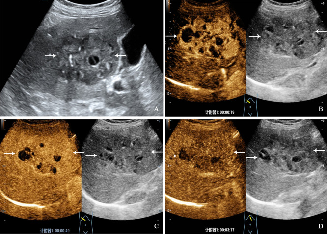
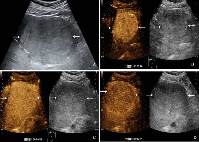
Results: PHNET was more common in women (57.1%, 8/14 cases), and NBNC-HCC was more common in men (75.0%, 21/28) (P=0.040). No significant differences were observed in etiology, tumor marker, and liver function between the two group (P>0.05). Conventional ultrasound revealed that the tumor size of PHNET (10.1 ± 4.7 cm) was larger than that of NBNC-HCC (5.9 ± 3.8 cm) (P=0.006). NBNC-HCC was predominantly hypoechoic, while the echogenicity of PHNET varied (P=0.001). On CEUS, 57.1% (8/14) of PHNETs showed heterogeneous hyperenhancement, whereas 77.0% (21/28) of NBNC-HCC presented homogeneous hyperenhancement (P=0.015). Furthermore, 35.7% (5/14) of PHNETs showed early washout (onset of washout <60 s), which was significantly different from that of NBNC-HCC (3.7%, 1/28) (P=0.005).

Conclusion: CEUS is helpful in discriminating between PHNET and NBNC-HCC. PHNETs mainly present as a single mass with a large size (>10 cm) in the liver. The CEUS showed that most PHNETs exhibited heterogeneous enhancement in the arterial phase, washout in the portal venous and late phases and early washout being more likely than NBNC-HCC. However, more imaging features need to be evaluated in a larger sample.

Keywords: contrast-enhanced ultrasound; hepatocellular carcinoma; liver; primary hepatic neuroendocrine tumor; ultrasonography.


Conflict of interest statement

The authors declare that the research was conducted in the absence of any commercial or financial relationships that could be construed as a potential conflict of interest.

Figures

Figure 1
Figure 1
The flowchart for inclusion of patients with primary hepatic neuroendocrine tumor in the study.
Figure 2
Figure 2
A 68-year-old woman with primary neuroendocrine tumor. The patient had no underlying liver disease. Conventional ultrasound showed that a mixed echoic tumor with focal calcification and largest diameter of 10.0 centimeters in anterior segment of the liver (A). In the arterial phase of contrast-enhanced ultrasound, the tumor showed heterogeneous hyperenhancement (B), and began washout before 60 seconds (C). hypoenhancement in the late phase (D).
Figure 3
Figure 3
A 32-year-old man with primary neuroendocrine tumor. The patient had no underlying liver disease. Conventional ultrasound showed that a slightly hyperechoic tumor with largest diameter of 10.3 centimeters in left liver lobe (A). In the arterial phase of contrast-enhanced ultrasound, the tumor showed homogeneous hyperenhancement (B), and began washout at 56 seconds (C), hypoenhancement in the late phase (D).
Figure 4
Figure 4
A 71-year-old man with hepatocellular carcinoma. The patient had no chronic hepatitis B and C Conventional ultrasound showed that a hypoechoic tumor with largest diameter of 3.8 centimeters in right liver lobe (A). In the arterial phase of contrast-enhanced ultrasound, the tumor showed homogeneous hyperenhancement (B), and isoenhancement in the portal venous phase (C), finally with slightly washout in the late phase (D).

References

    1. Shah MH, Goldner WS, Benson AB, Bergsland E, Blaszkowsky LS, Brock P, et al. . Neuroendocrine and adrenal tumors, version 2.2021, NCCN clinical practice guidelines in oncology. J Natl Compr Canc Netw (2021) 19(7):839–68. doi: 10.6004/jnccn.2021.0032 - DOI - PubMed
    1. Dasari A, Shen C, Halperin D, Zhao B, Zhou S, Xu Y, et al. . Trends in the incidence, prevalence, and survival outcomes in patients with neuroendocrine tumors in the united states. JAMA Oncol (2017) 3(10):1335–42. doi: 10.1001/jamaoncol.2017.0589 - DOI - PMC - PubMed
    1. Torbenson M, Venkatesh SK, Halfdanarson TR, Navin PJ, Kamath P, Erickson LA. Primary neuroendocrine tumors and primary neuroendocrine carcinomas of the liver: a proposal for a multidiscipline definition. Hum Pathol (2022) S0046-8177(22):00174–5. doi: 10.1016/j.humpath.2022.07.001 - DOI - PubMed
    1. Park CH, Chung JW, Jang SJ, Chung MJ, Bang S, Park SW, et al. . Clinical features and outcomes of primary hepatic neuroendocrine carcinomas. J Gastroenterol Hepatol (2012) 27(8):1306–11. doi: 10.1111/j.1440-1746.2012.07117.x - DOI - PubMed
    1. Huang J, Yu JQ, Sun JY. Computer tomography and magnetic resonance image manifestations of primary hepatic neuroendocrine cell carcinomas. Asian Pac J Cancer Prev (2014) 15(6):2759–64. doi: 10.7314/apjcp.2014.15.6.2759 - DOI - PubMed

LinkOut - more resources