Review
. 2022 Apr 15:2:809373.
doi: 10.3389/fradi.2022.809373. eCollection 2022.

Advanced MRI Protocols to Discriminate Glioma From Treatment Effects: State of the Art and Future Directions

Affiliations
Review

Advanced MRI Protocols to Discriminate Glioma From Treatment Effects: State of the Art and Future Directions

Dania G Malik et al. Front Radiol. .

Abstract

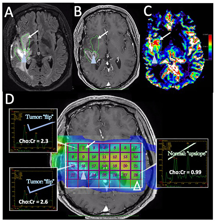
In the follow-up treatment of high-grade gliomas (HGGs), differentiating true tumor progression from treatment-related effects, such as pseudoprogression and radiation necrosis, presents an ongoing clinical challenge. Conventional MRI with and without intravenous contrast serves as the clinical benchmark for the posttreatment surveillance imaging of HGG. However, many advanced imaging techniques have shown promise in helping better delineate the findings in indeterminate scenarios, as posttreatment effects can often mimic true tumor progression on conventional imaging. These challenges are further confounded by the histologic admixture that can commonly occur between tumor growth and treatment-related effects within the posttreatment bed. This review discusses the current practices in the surveillance imaging of HGG and the role of advanced imaging techniques, including perfusion MRI and metabolic MRI.

Keywords: MRI; advanced; glioblastoma; metabolic; perfusion; post-treatment; response assessment.


Conflict of interest statement

KSw was co-founder of Precision Oncology Insights, US Patent US8571844B2. KSc: financial interest—Imaging Biometrics LLC; ownership interest—IQ-AQ Ltd; ownership interest—Prism Clinical Imaging, Inc. LH: Imaging Biometrics (Medical Advisory Board); Precision Oncology Insights (co-founder), Bayer Pharmaceutical (paid speaker), US Patent Number 10,909,675. The remaining authors declare that the research was conducted in the absence of any commercial or financial relationships that could be construed as a potential conflict of interest.

Figures

Figure 1
Figure 1
Posttreatment changes on Dynamic Susceptibility (DSC) MRI: A 58-year-old man with right frontal glioblastoma (GBM) status post resection and adjuvant radiation with concomitant temozolomide presented 6 months after resection with left facial droop and left-sided weakness. Initial CT and Contrast-enhanced MRI (CE-MRI) showed an increased mass effect and ill-defined enhancement around the resection cavity, indeterminant for recurrent tumor vs. posttreatment effects. (A) Axial postcontrast T1-weighted image demonstrates a linear enhancement around the right frontal resection cavity with more ill-defined nodular enhancement along the posteromedial margin (arrow). (B) Axial T2-weighted image shows T2 hyperintense signal and mass effect throughout the right frontal and parietal lobes. (C) DSC Fractional tumor burden (FTB) map derived from rCBV thresholds shows low FTB around the resection cavity favoring posttreatment changes (D) Stereotactic biopsies were performed and cross-registered retrospectively to areas of enhancement. Pathology demonstrated virtually entirely necrotic tissue with no viable tumor, concordant with the rCBV map.
Figure 2
Figure 2
Posttreatment changes on DSC-MRI: A 68-year-old woman with left frontal GBM with two prior surgical resections and adjuvant radiation, temozolomide, and lomustine therapy undergoing routine surveillance imaging. (A) CE-MRI 2 years after initial diagnosis demonstrated increased nodular enhancement along the resection cavity concerning recurrent tumor. (B) The DSC FTB map derived from relative cerebral blood volume (rCBV) thresholds demonstrate a small area of high FTB centrally (arrowhead) suggesting tumor recurrence, but the majority of the surrounding tissue had low rCBV consistent with posttreatment effect. (C) Stereotactic biopsies were performed and cross-registered with areas of enhancement, but only corresponded with low rCBV regions. Pathology showed scant atypical cells in a background of extensive therapy-related changes, compatible with the rCBV map. No viable GBM was identified. Unfortunately, the central portion of the tumor was not sampled and cross-registration with the DSC images at the time of biopsy may have been helpful in fully characterizing the lesion pathologically.
Figure 3
Figure 3
Recurrent tumor on DSC-MRI: A 27-year-old man with right temporoparietal GBM with prior surgical resection with carmustine wafer placement and adjuvant radiation/temozolomide. Five months after surgical resection, the patient experienced clinical deterioration with signs of increased intracranial pressure with clinical concern for tumor progression vs. pseudoprogression. His symptoms did improve with steroid therapy. (A) Axial postcontrast T1 weighted image shows a large area of nodular and ring-like enhancement along the medial margin of the resection cavity (arrow). (B) The DSC FTB map derived from rCBV thresholds demonstrates high FTB centrally in the area of nodular enhancement consistent with the tumor. (C) The patient underwent surgical debulking, and the stereotactic biopsy at the site of the elevated rCBV (circular marker) was positive for 80% recurrent viable tumor.
Figure 4
Figure 4
Percentage signal recovery in PTRE and recurrent tumor: A 68-year-old woman with left frontal GBM with two prior surgical resections and adjuvant radiation, temozolomide, and lomustine therapy undergoing routine surveillance imaging. DSC was performed due to concern for tumor recurrence (refer to Figure 2). The FTB map shows peripheral low rCBV (blue) consistent with PTRE which was confirmed on stereotactic biopsy. The central component with higher rCBV (yellow) was presumed to have a recurrent tumor and resected, but not confirmed with stereotactic biopsy. Correlating PSR maps and signal intensity-time curves are provided to demonstrate a high PSR of essentially 100% for the peripheral region consistent with the PTRE and a lower PSR of 58% for the central region that was presumed to have a recurrent tumor.
Figure 5
Figure 5
Disease recurrence on DSC-MRI and MRS: A 50-year-old man with right temporal WHO grade III anaplastic oligodendroglioma status post three surgical resections, radiation therapy, and chemotherapy with temozolomide, procarbazine, and lomustine was found to have signs of recurrence on surveillance CE-MRI. (A) Axial FLAIR image superior to the resection cavity shows prominent increased FLAIR signal posteriorly (blue arrowhead) and less intense, but more rounded FLAIR signal anteriorly (arrow). (B) Axial post-contrast T1-weighted image demonstrates heterogenous enhancement of the posterior region (blue arrowhead) with no significant enhancement in the anterior region (arrow) (C) The DSC rCBV map shows increased perfusion of the anterior nonenhancing/FLAIR hyperintense region (arrow) but heterogeneous areas of both increased and decreased perfusion in the enhancing posterior region (arrowhead). (D) Magnetic resonance spectroscopy (MRS) demonstrates elevated Cho:Cr ratios in both the anterior nonenhancing (arrow, ratio = 2.3) and the posterior enhancing (solid blue arrowhead, ratio = 2.6) regions. Normal ratios are shown on the contralateral brain (open arrowhead, Cho:Cr ratio = 0.99) for comparison, showing the normal upslope vs. the “Cho-to-Cr flip” seen with the tumor. Both sites with elevated Cho:Cr ratio was concerning for disease progression. The heterogeneous rCBV of the posterior enhancing region was attributed to a combination of disease recurrence and necrotic change. The patient was treated with repeat proton-beam radiation and temozolomide with improvement in the enhancement and FLAIR signal abnormality on subsequent MRI (not shown).

References

    1. National Comprehensive Cancer Network . Central Nervous System Cancers. (Version 2.2021). Available online at: https://www.nccn.org/professionals/physician_gls/pdf/cns.pdf (accessed October 7, 2021).
    1. Ellingson BM, Bendszus M, Boxerman J, Barboriak D, Erickson BJ, Smits M, et al. . Consensus recommendations for a standardized brain tumor imaging protocol in clinical trials. Neuro Oncol. (2015) 17:1188–98. 10.1093/neuonc/nov095 - DOI - PMC - PubMed
    1. Verma N, Cowperthwaite MC, Burnett MG, Markey MK. Differentiating tumor recurrence from treatment necrosis: a review of neuro-oncologic imaging strategies. Neuro Oncol. (2013) 15:515–34. 10.1093/neuonc/nos307 - DOI - PMC - PubMed
    1. Walker MD, Alexander E Jr, Hunt WE, MacCarty CS, Mahaley MS Jr, Mealey J Jr, et al. . Evaluation of BCNU and/or radiotherapy in the treatment of anaplastic gliomas. A cooperative clinical trial: a cooperative clinical trial. J Neurosurg. (1978) 49:333–43. 10.3171/jns.1978.49.3.0333 - DOI - PubMed
    1. Stupp R, Mason WP, van den Bent MJ, Weller M, Fisher B, Taphoorn MJB, et al. . Radiotherapy plus concomitant and adjuvant temozolomide for glioblastoma. N Engl J Med. (2005) 352:987–96. 10.1056/NEJMoa043330 - DOI - PubMed

LinkOut - more resources