Review
. 2023 Nov;96(1151):20230232.
doi: 10.1259/bjr.20230232. Epub 2023 Jul 26.

Insights into acute mesenteric ischaemia: an up-to-date, evidence-based review from a mesenteric stroke centre unit

Affiliations
Review

Insights into acute mesenteric ischaemia: an up-to-date, evidence-based review from a mesenteric stroke centre unit

Lorenzo Garzelli et al. Br J Radiol. 2023 Nov.

Abstract

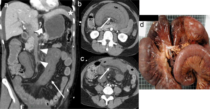
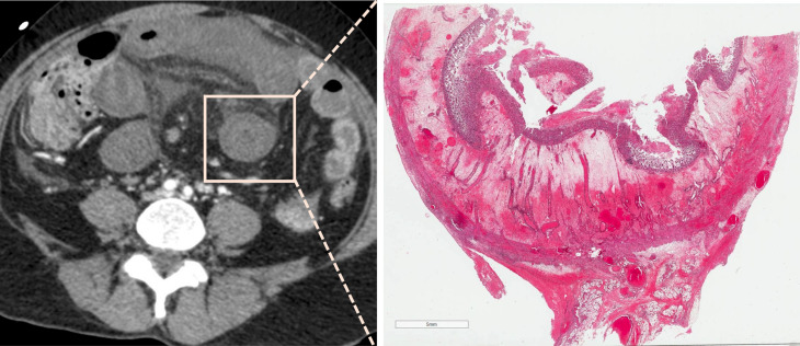
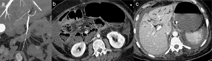
Radiologists play a central role in the diagnostic and prognostic evaluation of patients with acute mesenteric ischaemia (AMI). Unfortunately, more than half of AMI patients undergo imaging with no prior suspicion of AMI, making identifying this disease even more difficult. A confirmed diagnosis of AMI is ideally made with dynamic contrast-enhanced CT but the diagnosis may be made on portal-venous phase images in appropriate clinical settings. AMI is diagnosed on CT based on the identification of vascular impairment and bowel ischaemic injury with no other cause. Moreover, radiologists must evaluate the probability of bowel necrosis, which will influence the treatment options.AMI is usually separated into different entities: arterial, venous, non-occlusive and ischaemic colitis. Arterial AMI can be occlusive or stenotic, the dominant causes being atherothrombosis, embolism and isolated superior mesenteric artery (SMA) dissection. The main finding in the bowel is decreased wall enhancement, and necrosis can be suspected when dilatation >25 mm is identified. Venous AMI is related to superior mesenteric vein (SMV) thrombosis as a result of a thrombophilic state (acquired or inherited), local injury (cancer, inflammation or trauma) or underlying SMV insufficiency. The dominant features in the bowel are hypoattenuating wall thickening with submucosal oedema. Decreased enhancement of the involved bowel suggests necrosis. Non-occlusive mesenteric ischaemia (NOMI) is related to impaired SMA flow following global hypoperfusion associated with low-flow states. There are numerous findings in the bowel characterised by diffuse extension. An absence of bowel enhancement and a thin bowel wall suggest necrosis in NOMI. Finally, ischaemic colitis is a sub-entity of arterial AMI and reflects localised colon ischaemia-reperfusion injury. The main CT finding is a thickened colon wall with fat stranding, which seems to be unrelated to SMA or inferior mesenteric artery lesions. A precise identification and description of vascular lesions, bowel involvement and features associated with transmural necrosis is needed to determine patient treatment and outcome.


Figures

Figure 1.
Figure 1.
Illustration of the superior mesenteric artery segmentation
Figure 2.
Figure 2.
Example of decreased bowel wall enhancement in early arterial occlusive acute mesenteric ischaemia. Panel A shows a non-dilated bowel, but some segments are hypoenhancing compared to others (arrows), with clear demarcation between ischaemic and non-ischaemic segments. Different bowel segments can be seen on panel B: mildly hypoenhancing non-dilated loop suggesting ischaemic injury (left side), a normal enhancing non-dilated loop free from ischaemic involvement (middle) and a hypoenhancing slightly dilated loop suggesting more advanced ischaemic, but still non-necrotic, lesions (right side).
Figure 3.
Figure 3.
87-year-old female patient with early acute occlusive arterial mesenteric ischaemia (AMI). Image A shows occlusion of the distal segment of the superior mesenteric artery (SMA, segment S3) by a clot (arrow) with patent proximal and middle SMA (segment S1 and S2) and ileocolic artery (arrowheads). Images B and C show decreased enhancement of a non-dilated ileal wall (arrows) with normal enhancement of the adjacent small bowel (star). These features suggest early non-necrotic AMI. However, the patient underwent ileocecal resection in another institution. The pathologic examination showed a yellowish mucosa with a tiger stripe appearance but with a normal outer layer (macroscopic appearance D) and no transmural necrosis on pathology. This patient had a Clichy score of 0/3 (small bowel diameter <25 mm, lactate <2 mmol l−1 and absence of organ failure) and could have avoided resection if properly revascularised.
Figure 4.
Figure 4.
Example of a delayed diagnosis resulting in transmural necrosis in a 56-year-old male patient. This patient presented in the emergency department with marked and intense abdominal pain. Because of acute kidney injury, he underwent non-contrast abdominal computed tomography (to rule out a perforated ulcer), showing isolated dilatation of the ileum (A). He was admitted for observation, but he presented with sepsis and lactic acidosis 12 hours later. He underwent a contrast-enhanced computed tomography (B, portal venous phase), showing a hypo-enhancing dilated ileum and occlusion of the superior mesenteric artery (not depicted here). Two and a half meters of small bowel were resected. The patient is alive but now has short bowel syndrome.
Figure 5.
Figure 5.
An 86-year-old female patient presented to the emergency department with marked abdominal pain requiring morphine and elevated serum lactate at 3.2 mmol l−1. Image A shows an embolus of the middle and distal segments of the superior mesenteric artery (segment S2 and S3, arrow). Unenhanced computed tomography (B) showed dilatation of the ileum (arrow) and portal-venous phase (C) showed decreased bowel enhancement. This patient underwent bowel resection because of extensive macroscopic signs of necrosis (D) with 1 m of small bowel left in place. Transmural necrosis was found in microscopic evaluation.
Figure 6.
Figure 6.
Acute embolic occlusive arterial mesenteric ischaemia in a 60-year-old male patient. Images A and B show an embolus of the superior mesenteric artery with decreased enhancement of the ileum (arrow). This patient underwent open surgical thrombectomy resulting in complete revascularisation. Images C and D show the follow-up CT scan with a patent SMA and bowel wall thickening (with associated hypoattenuating oedema of the submucosa and hyperenhancement of the mucosa) of the ileum (arrow) suggesting a reperfusion injury.
Figure 7.
Figure 7.
Example of acute superior mesenteric venous thrombosis in a 48-year-old female patient with abdominal pain. Images A and B show complete thrombosis of the superior mesenteric vein with parietal enhancement and adjacent fat stranding (arrows) corresponding to thrombosis. In images C and D, the bowel shows no features of ischaemic injury (i.e., normal enhancement, no wall thickening).
Figure 8.
Figure 8.
Necrotic acute venous mesenteric ischaemia in a 59-year-old female patient who presented to the emergency department for marked abdominal pain requiring morphine. Images A, B and C show hypoattenuating bowel wall thickening with a multiple-layer appearance, suggesting submucosal bowel oedema (arrow) with complete superior mesenteric vein thrombosis (A, arrowhead) and ascites (A, star). In addition, the involved bowel loops showed decreased enhancement suggesting necrosis. Image D shows the macroscopic analysis with a dark brown small bowel and congestive mesentery with transmural necrosis plus haemorrhage on microscopy.
Figure 9.
Figure 9.
Non-necrotic non-occlusive mesenteric ischaemia in a 69-year-old male patient after aortocoronary bypass. Biological laboratory tests showed a lactate concentration of 5 mmol l−1, a prothrombin rate of 79% and a bicarbonate concentration of 22 mmol l−1. Image A shows a diffuse spasm of the superior mesenteric artery. Image B shows a small bowel dilatation with persistent wall enhancement, and image C shows portal venous gas (secondary to bowel wall pneumatosis, not shown). Despite the presence of pneumatosis, this patient had a low risk of necrosis according to the NOMI-ITN score. The patient underwent a laparotomy that did not reveal any bowel necrosis.

References

    1. Schoots IG, Koffeman GI, Legemate DA, Levi M, van Gulik TM. Systematic review of survival after acute mesenteric ischaemia according to disease Aetiology. Br J Surg 2004; 91: 17–27. doi: 10.1002/bjs.4459 - DOI - PubMed
    1. Lemma A, Tolonen M, Vikatmaa P, Mentula P, Kantonen I, But A, et al. . Editor’s choice - epidemiology, diagnostics, and outcomes of acute occlusive arterial mesenteric ischaemia: A population based study. Eur J Vasc Endovasc Surg 2022; 64: 646–53. doi: 10.1016/j.ejvs.2022.07.006 - DOI - PubMed
    1. Cokkinis AJ. Mesenteric vascular occlusion. Southern Medical Journal 1926; 19: 655. doi: 10.1097/00007611-192608000-00028 - DOI
    1. Nuzzo A, Maggiori L, Ronot M, Becq A, Plessier A, Gault N, et al. . Predictive factors of intestinal necrosis in acute mesenteric ischemia: prospective study from an intestinal stroke center. Am J Gastroenterol 2017; 112: 597–605. doi: 10.1038/ajg.2017.38 - DOI - PubMed
    1. Najdawi M, Garzelli L, Nuzzo A, Huguet A, Raynaud L, Paulatto L, et al. . Endovascular Revascularization of acute arterial mesenteric ischemia: report of a 3-year experience from an intestinal stroke center unit. Eur Radiol 2022; 32: 5606–5615. doi: 10.1007/s00330-022-08660-3 - DOI - PubMed