. 2023 Nov 1;22(11):1248-1260.
doi: 10.1158/1535-7163.MCT-23-0031.

Preclinical Development of Brain Permeable ERβ Agonist for the Treatment of Glioblastoma

Affiliations

Preclinical Development of Brain Permeable ERβ Agonist for the Treatment of Glioblastoma

Uday P Pratap et al. Mol Cancer Ther. .

Abstract

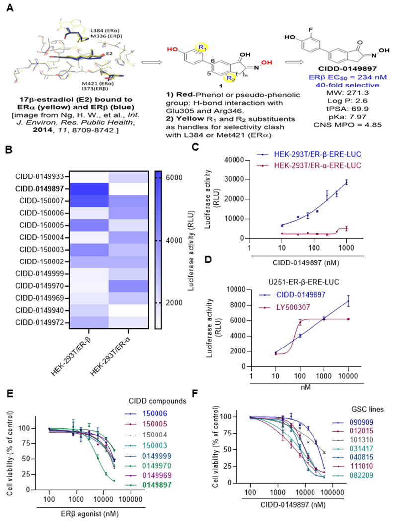
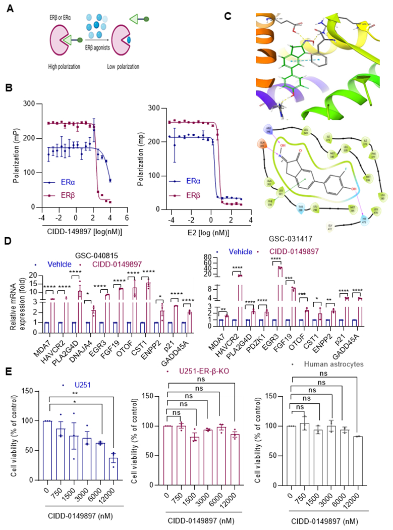
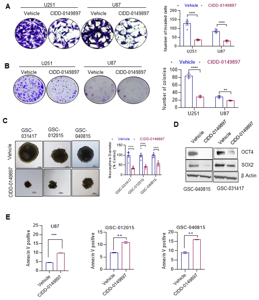
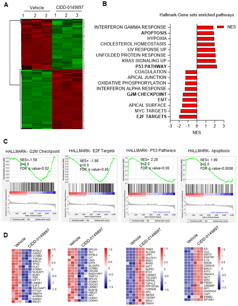
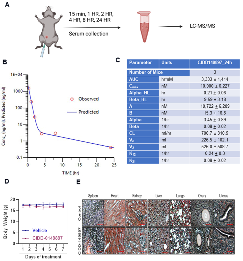
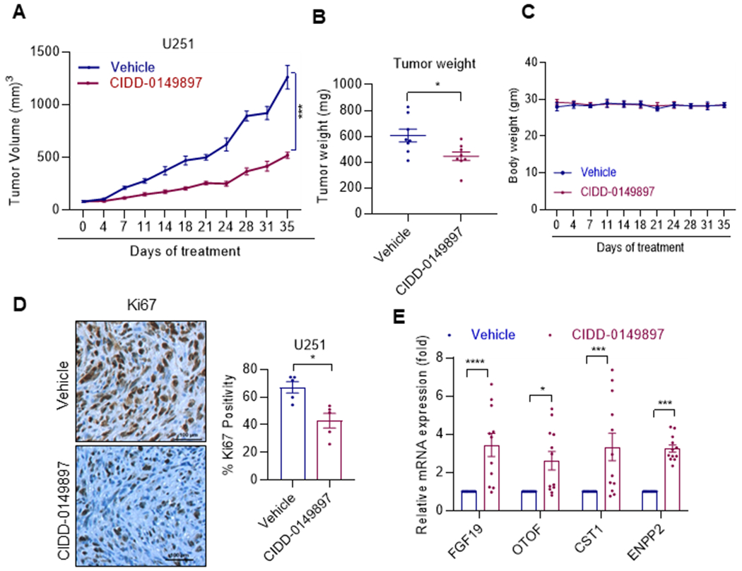
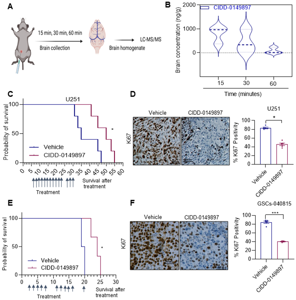
Glioblastoma (GBM) is the most prevalent and aggressive type of adult brain tumors with low 5-year overall survival rates. Epidemiologic data suggest that estrogen may decrease brain tumor growth, and estrogen receptor beta (ERβ) has been demonstrated to exert antitumor functions in GBM. The lack of potent, selective, and brain permeable ERβ agonist to promote its antitumor action is limiting the therapeutic promise of ERβ. In this study, we discovered that Indanone and tetralone-keto or hydroxyl oximes are a new class of ERβ agonists. Because of its high activity in ERβ reporter assays, specific binding to ERβ in polar screen assays, and potent growth inhibitory activity in GBM cells, CIDD-0149897 was discovered as a possible hit by screening a library of compounds. CIDD-0149897 is more selective for ERβ than ERα (40-fold). Treatment with CIDD-0149897 markedly reduced GBM cell viability with an IC50 of ∼7 to 15 μmol/L, while having little to no effect on ERβ-KO cells and normal human astrocytes. Further, CIDD-0149897 treatment enhanced expression of known ERβ target genes and promoted apoptosis in established and patient-derived GSC models. Pharmacokinetic studies confirmed that CIDD-0149897 has systemic exposure, and good bioavailability in the brain. Mice tolerated daily intraperitoneal treatment of CIDD-0149897 (50 mg/kg) with a 7-day repeat dosage with no toxicity. In addition, CIDD-0149897 treatment significantly decreased tumor growth in U251 xenograft model and extended the survival of orthotopic GBM tumor-bearing mice. Collectively, these findings pointed to CIDD-0149897 as a new class of ERβ agonist, offering patients with GBM a potential means of improving survival.


Conflict of interest statement

Competing interests: UT System filed a patent application on ER beta agonists

Figures

Figure 1.
Figure 1.
Identification of CIDD-0149897 as novel ERβ agonist. A, Schematic representation of ERβ ligand design strategy and the structure of CIDD-0149897. B HEK293T model cells stably expressing ERβ or ERα along with ERE-Luc reporter was used for initial screening of compounds testing for ER activity (n=3). Heat map showing ERβ and ERα reporter activity of selective compounds (n=3). C, HEK293Tcells expressing ERβ or ERα were treated with various doses of CIDD-0149897 and ERE-Luc reporter activity was measured after 24 hours (n=2). D, U251 GBM cells expressing ERβ were treated with indicated doses of CIDD-0149897 or LY500307 and after 24 h, ERE-Luc reporter activity was measured (n=3). E. Effect of increasing doses of selected ERβ agonists on the cell viability of U251 GBM model cells expressing ERβ was measured using the MTT cell viability assay (n=3). F, Effect of increasing doses of CIDD-0149897 on the cell viability of indicated patients derived GSCs was measured using CellTiter Glo assay (n=3). Data are presented as mean ± SEM.
Figure 2.
Figure 2.
Target specificity of CIDD-0149897. A, Schematic of polar screen ER binding assay. B, Specificity of E2 (left panel) and CIDD-0149897 (right panel) to ERα and ERβ determined using polar screen ER competitive binding assays. Fluorescence polarization is measured and shown as millipolarization (mP) (n=2) C, Induced fit docking pose of CIDD-0149897 in the ligand binding site of PDB structure 1X7B. (Top panel) 3D rendering of docking pose indicating hydrogen bonding (yellow) and pi-stacking (cyan) interactions. (Bottom panel) 2D representation of ligand interactions. D, Effect of CIDD-0149897 treatment (10 μM, 72h) on ERβ targeted genes was measured using RT-qPCR analysis in GSC-040815, GSC-031417cells (n=3). E, Effect of CIDD-0149897 on normal human astrocytes as well as on WT and ERβ-KO U251 GBM cells was measured using the MTT cell viability assay (n=3). Data are presented as mean ± SEM * p<0.05, ** p<0.01, *** p<0.001, ****p<0.0001. In D, p-values were calculated using two-way ANOVA. In E, p-values were calculated using one-way ANOVA.
Figure 3.
Figure 3.
CIDD-0149897 decreases invasion, colony formation, neurosphere formation, stemness and promotes apoptosis of GBM cells. A, Effect of CIDD-0149897 (10μM, 22h) on cell invasion of U251 and U87 GBM model cells was determined using Matrigel invasion chamber assays (n=3). The number of invaded cells in five randomly selected fields was quantitated, and representative photos of invading cells are displayed. B, Effect of CIDD-0149897 (10 μM) on cell survival was measured using colony formation assays. C, Effect of CIDD-0149897 (20 μM) on neurosphere formation was measured in GSC-031417, GSC-012015, and GSC-040815 cells (n=3). Single cell suspension of GSCs were seeded in 24 well plates (100 cells/well) and treated with vehicle or CIDD-0149897 and after 7 days neurosphere were photographed and quantitated. GSCs were treated with vehicle or CIDD-0149897 and the expression levels of stemness genes were determined using western blotting (D). E, Effect of CIDD-0149897 (20 μM) on apoptosis was measured by Annexin V staining in GBM model cells. Data are presented as mean ± SEM. ** p<0.01, *** p<0.001, ****p<0.0001. In A, B and C, p-values were calculated using two-way ANOVA. In E, p-values were calculated using the t test.
Figure 4.
Figure 4.
Global analysis of transcriptional changes altered by CIDD-0149897 treatment. A, DEGs identified from RNA-seq data are displayed in Heatmap. B, CIDD-0149897 regulated genes were subjected to GSEA and top positively or negatively enriched gene sets from Hallmark pathways were shown. C, GSEA enrichment plots of the apoptosis, p53, E2F, and G2/M checkpoint pathways were shown. D, heatmap of the selected genes related to the above indicated pathways are shown.
Figure 5.
Figure 5.
PK and toxicity analyses of CIDD-0149897. A, schematic of PK study design. B, C57/BL6 female mice (n=3) were administered with CIDD-0149897 (20mg/kg/single dose) intraperitoneally, and PK of the compounds at different time points was analyzed in blood. C, Summary of PK data for CIDD-0149897. D, C57BL6 mice were treated with CIDD-0149897 (50 mg/kg/i.p./day) or vehicle treatment for seven days and body weights were recorded. E, Histologic architecture of multiple organs in C57BL/6 mice were evaluated using H&E staining. Data are presented as mean ± SEM.
Figure 6.
Figure 6.
CIDD-0149897 inhibits the growth of GBM xenograft tumors. A, U251 xenografts (n=4) were treated with vehicle or CIDD-014897 (20mg/kg/i. p./5 days/week). B, Tumor volumes are shown in the graph. C, Body weights of vehicle and CIDD-014897 treated mice are shown. D, Ki-67 expression as a marker of proliferation was analyzed by IHC and quantitated. E, Status of ERβ induced target genes were measured by using RT-qPCR analysis (n=3). * p<0.05, *** p<0.001, ****p<0.0001. Data are presented as mean ± SEM. In A, p-values were calculated using linear mixed effect model. In B and D, p-values were calculated using the t test and in E, p value were calculated using two-way ANOVA.
Figure 7.
Figure 7.
CIDD-0149897 has BBB permeability and therapeutic effects on orthotopic xenografts of GBM. A, Schematic of design used for studying brain tissue concentration of CIDD-0149897. B, C57BL/6 mice (female, n=3) were treated with a single dose of CIDD-0149897 (50 mg/kg/i.p./day) and the brain tissue concentrations were determined at indicated times. C, Mice were implanted with U251 cells orthotopically in the right cerebrum. After the tumor establishment, mice (n=5) were treated with vehicle (control) or CIDD-0149897 (50 mg/kg/i.p./day) and the survival of the mice was plotted using Kaplan-Meier curve. E, Patient derived GSC-040815- cells were implanted orthotopically in SCID mice, treated with vehicle (control) or CIDD-014987 (50 mg/kg/i.p.) for 11 days as indicated and the survival of the mice was plotted using Kaplan-Meier curve (n = 5). D, F, Ki-67 expression as a marker of proliferation was analyzed by IHC and quantitated. * p<0.05, *** p<0.001. Data are presented as mean ± SEM. In D and E, p-values were calculated using the t test. In C and E, p-values were calculated using Log-rank (Mantel-Cox) test.

References

    1. Ohgaki H, Kleihues P. Genetic alterations and signaling pathways in the evolution of gliomas. Cancer Sci 2009;100(12):2235–41 doi 10.1111/j.1349-7006.2009.01308.x. - DOI - PMC - PubMed
    1. Omuro A, DeAngelis LM. Glioblastoma and other malignant gliomas: a clinical review. JAMA 2013;310(17):1842–50 doi 10.1001/jama.2013.280319. - DOI - PubMed
    1. Tykocki T, Eltayeb M. Ten-year survival in glioblastoma. A systematic review. J Clin Neurosci 2018;54:7–13 doi 10.1016/j.jocn.2018.05.002. - DOI - PubMed
    1. Stupp R, Taillibert S, Kanner A, Read W, Steinberg D, Lhermitte B, et al. Effect of Tumor-Treating Fields Plus Maintenance Temozolomide vs Maintenance Temozolomide Alone on Survival in Patients With Glioblastoma: A Randomized Clinical Trial. JAMA 2017;318(23):2306–16 doi 10.1001/jama.2017.18718. - DOI - PMC - PubMed
    1. Johnson DR, O’Neill BP. Glioblastoma survival in the United States before and during the temozolomide era. Journal of neuro-oncology 2012;107(2):359–64 doi 10.1007/s11060-011-0749-4. - DOI - PubMed

Publication types

Associated data