. 2023 Aug 3;40(8):msad171.
doi: 10.1093/molbev/msad171.

Whole Genome Duplication and Gene Evolution in the Hyperdiverse Venomous Gastropods

Affiliations

Whole Genome Duplication and Gene Evolution in the Hyperdiverse Venomous Gastropods

Sarah Farhat et al. Mol Biol Evol. .

Abstract

The diversity of venomous organisms and the toxins they produce have been increasingly investigated, but taxonomic bias remains important. Neogastropods, a group of marine predators representing almost 22% of the known gastropod diversity, evolved a wide range of feeding strategies, including the production of toxins to subdue their preys. However, whether the diversity of these compounds is at the origin of the hyperdiversification of the group and how genome evolution may correlate with both the compounds and species diversities remain understudied. Among the available gastropods genomes, only eight, with uneven quality assemblies, belong to neogastropods. Here, we generated chromosome-level assemblies of two species belonging to the Tonnoidea and Muricoidea superfamilies (Monoplex corrugatus and Stramonita haemastoma). The two obtained high-quality genomes had 3 and 2.2 Gb, respectively, and 92-89% of the total assembly conformed 35 pseudochromosomes in each species. Through the analysis of syntenic blocks, Hox gene cluster duplication, and synonymous substitutions distribution pattern, we inferred the occurrence of a whole genome duplication event in both genomes. As these species are known to release venom, toxins were annotated in both genomes, but few of them were found in homologous chromosomes. A comparison of the expression of ohnolog genes (using transcriptomes from osphradium and salivary glands in S. haemastoma), where both copies were differentially expressed, showed that most of them had similar expression profiles. The high quality of these genomes makes them valuable reference in their respective taxa, facilitating the identification of genome-level processes at the origin of their evolutionary success.

Keywords: genomes; neofunctionalization; neogastropods; venom; whole-genome duplication.


Figures

Fig. 1.
Fig. 1.
Schematic representation of the phylogenetic relationships between the species compared in this study. Simplified tree of the four Caenogastropoda species studied in this work. Numbers for each pair correspond to the number of orthologs detected between the two species (top) and the number of syntenic regions detected between the two genomes, all chromosomes considered (bottom). The three first pictures credits: MNHN CC BY 4.0. The picture of P. canaliculata was taken from Ng et al. 2017.
Fig. 2.
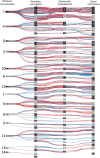
Fig. 2.
Syntenic blocks in Caenogastropoda. Representation of the pseudochromosomes (black and gray rectangles with their pseudochromosome numbers) of the four Caenogastropoda generated using Synvisio (Bandi and Gutwin 2020). Each line represents a syntenic block in reverse or in forward orientation. Only links between side-by-side species are represented by full lines, whereas the dotted lines represent links between not juxtaposed species (no synteny detected between the two juxtaposed pseudochromosomes).
Fig. 3.
Fig. 3.
(A) Orthologs, paralogs, and ohnologs. The rectangle on a line represents a gene on a chromosome. Lines from species 1 to species 2 identify orthologs; the upper curved line defines paralogs (in tandem on the same chromosome); the lower curved line identify ohnologs (in two homologous chromosomes within a single species). (B) Ks density plot. Distribution of synonymous divergence between pairs of paralogs (first three lines, Conus, Monoplex and Stramonita) and orthologs (for all remaining lines). The first peak represents the newest events of duplication of each species (Conus ventricosus, Monoplex corrugatus, and Stramonita haemastoma); the middle peak corresponds to the whole genome duplication event; and the third peak represents the point of divergence between P. canaliculata and the three other species.
Fig. 4.
Fig. 4.
Hox genes clusters. (A) Representation of the gene order of Hox genes; colored boxes with the arrow indicating the gene direction on the pseudochromosome (Chr) or superscaffold (Sc) represented by a line, as detected during the annotation. Star marks species for which data were retrieved from literature (Liu et al. 2020; Pardos-Blas et al. 2021). (B) Tree representing the phylogenetic relationships between all the sequences of the analysis; color dots correspond to the species as listed on the (A) part of the figure.
Fig. 5.
Fig. 5.
Synteny block and multiple alignment of toxins similar to conotoxins. (A) Representation of syntenic blocks (light shadow) on each superscaffold/pseudochromosome and each species with putative toxins from group 4 (A1) and 1 (A2). Each horizontal line of a superscaffold/pseudochromosome represents one species. Red blocks are the detected toxins, black blocks are the genes found in the syntenic region, and green blocks are ortholog genes found in synteny (indicated by green lines). A toxin from group 1 found in Monoplex corrugatus (scaffold 2) was found in synteny with Pomacea canaliculata in a region that also shares a syntenic block with scaffold 1 of M. corrugatus close to the region of the paralog toxin. (B) Multiple alignment of group 4 (B1) and group 1 (B2) sequences (supplementary table S3, Supplementary Material online) manually annotated and found in a syntenic block with the addition of two conotoxins from Conus betulinus and C. magus, generated using MUSCLE v.3.8 (Edgar 2004).

References

    1. Akondi KB, Muttenthaler M, Dutertre S, Kaas Q, Craik DJ, Lewis RJ, Alewood PF. 2014. Discovery, synthesis, and structure–activity relationships of conotoxins. Chem Rev. 114:5815–5847. - PMC - PubMed
    1. Almeida DD, Viala VL, Nachtigall PG, Broe M, Gibbs HL, Serrano SMT, Moura-da-Silva AM, Ho PL, Nishiyama-Jr MY, Junqueira-de-Azevedo ILM. 2021. Tracking the recruitment and evolution of snake toxins using the evolutionary context provided by the Bothrops jararaca genome. Proc Natl Acad Sci. 118:e2015159118. - PMC - PubMed
    1. Anders S, Huber W. 2010. Differential expression analysis for sequence count data. Genome Biol. 11:R106. - PMC - PubMed
    1. Arbuckle K. 2018. Phylogenetic comparative methods can provide important insights into the evolution of toxic weaponry. Toxins (Basel). 10:518. - PMC - PubMed
    1. Balaji RA, Ohtake A, Sato K, Gopalakrishnakone P, Kini RM, Seow KT, Bay B-H. 2000. λ-Conotoxins, a new family of conotoxins with unique disulfide pattern and protein folding. J Biol Chem. 275:39516–39522. - PubMed

Publication types