. 2023 Jul 26;14(7):467.
doi: 10.1038/s41419-023-05997-5.

Nitric oxide-induced ribosome collision activates ribosomal surveillance mechanisms

Affiliations

Nitric oxide-induced ribosome collision activates ribosomal surveillance mechanisms

Laura Ryder et al. Cell Death Dis. .

Abstract

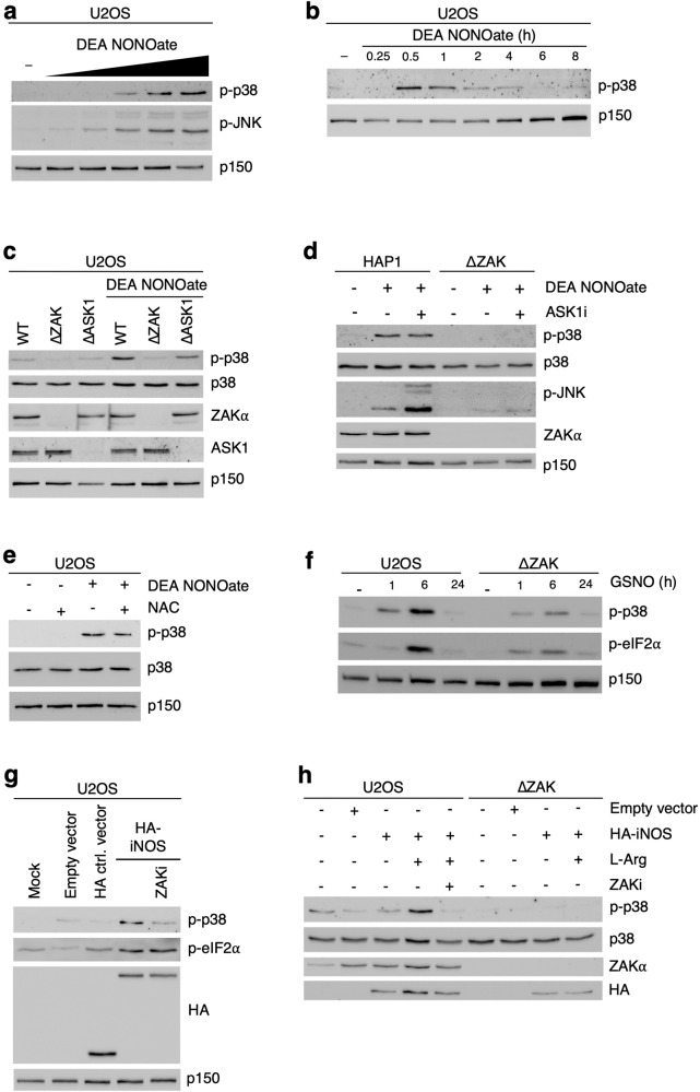
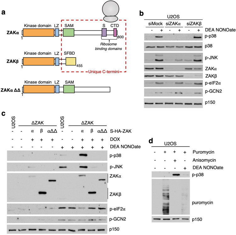
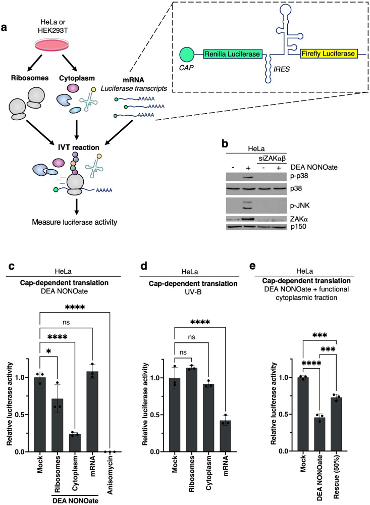
Impairment of protein translation can cause stalling and collision of ribosomes and is a signal for the activation of ribosomal surveillance and rescue pathways. Despite clear evidence that ribosome collision occurs stochastically at a cellular and organismal level, physiologically relevant sources of such aberrations are poorly understood. Here we show that a burst of the cellular signaling molecule nitric oxide (NO) reduces translational activity and causes ribosome collision in human cell lines. This is accompanied by activation of the ribotoxic stress response, resulting in ZAKα-mediated activation of p38 and JNK kinases. In addition, NO production is associated with ZNF598-mediated ubiquitination of the ribosomal protein RPS10 and GCN2-mediated activation of the integrated stress response, which are well-described responses to the collision of ribosomes. In sum, our work implicates a novel role of NO as an inducer of ribosome collision and activation of ribosomal surveillance mechanisms in human cells.


Conflict of interest statement

The authors declare no competing interests.

Figures

Fig. 1
Fig. 1. Nitric oxide activates p38 and JNK in a ZAK-dependent manner.
a U2OS cells were treated with increasing concentrations of DEA NONOate (100 μM, 250 μM, 500 μM, 750 μM, 1000 μM – 1 h). Lysates were analyzed by immunoblotting with the indicated antibodies. b U2OS cells were treated with DEA NONOate (750 μM) for the indicated times. Lysates were analyzed as in (a). c U2OS cells deleted for ZAK (ΔZAK) or ASK1 (ΔASK1) were treated with DEA NONOate (750 μM – 1 h). Lysates were analyzed as in (a). d HAP1 WT and ΔZAK cells were pretreated with ASK1 inhibitor (2 μM – 30 min) prior to treatment with DEA NONOate (750 μM – 1 h). Lysates were analyzed as in (a). e U2OS cells were pretreated with N-acetyl-L-cysteine (NAC, 10 mM – 1 h) followed by DEA NONOate treatment (750 μM – 1 h). Lysates were analyzed as in (a). f U2OS WT and ΔZAK cells were treated with S-Nitrosoglutathione (GSNO, 2 mM) for the indicated times. Lysates were analyzed as in (a). g U2OS cells were transfected either with empty vector or strep-HA-iNOS and treated with ZAK inhibitor (2 μM – 1 h). Lysates were analyzed as in (a). h U2OS WT and ΔZAK cells were transfected with empty vector or strep-HA-iNOS and supplemented with L-arginine (10 mM – 20 h) and ZAK inhibitor (2 μM – 1 h). Lysates were analyzed as in (a).
Fig. 2
Fig. 2. Nitric oxide donors inhibit translation and activate the ribotoxic stress response.
a Schematic of ZAK protein isoforms and the ZAKα ΔSΔCTD mutant. LZ, Leucine Zipper; SAM, Sterile Alpha-Motif; S, Sensor Domain; CTD, C-Terminal Domain; SFBD, Stress Fiber Binding Domain. b U2OS cells were mock-transfected or transfected with siRNAs targeting the α or the β isoform of ZAK. Cells were treated with DEA NONOate (750 μM – 1 h). Lysates were analyzed by immunoblotting with the indicated antibodies. c U2OS cells, ΔZAK and ΔZAK cells were rescued with WT ZAKα or ZAKβ, and a mutated form (αΔΔ) of ZAKα were treated with DEA NONOate (750 μM – 1 h). Lysates were analyzed as in (b). d U2OS cells were treated with DEA NONOate (750 μM – 1 h) or anisomycin (1 μg/ml – 1 h) followed by treatment with puromycin (10 μg/mL - 10 min). Lysates were analyzed as in (b).
Fig. 3
Fig. 3. Soluble translation factors are sensitive to nitrosative damage.
a Schematic of tri-partite in vitro translation (IVT) assay. Ribosomes and a ribosome-depleted cytoplasmic fraction were isolated from cultured HeLa or HEK293 cells and combined with in vitro transcribed luciferase mRNA for IVT. The three fractions can be individually treated before combination. b HeLa cells were mock-transfected or transfected with siRNAs targeting both isoforms (α and β) of ZAK. Cells were treated with DEA NONOate (750 μM – 1 h). Lysates were analyzed by immunoblotting with the indicated antibodies. c HeLa cell fractions from (a) were individually treated with DEA NONOate (1 mM – 10 min) prior to IVT (30 min, 37 °C). Anisomycin (1 μg/ml) was added directly into the full IVT mix as a positive control for translational inhibition. Cap-dependent translation efficiency in the combined reaction was determined by luciferase assay. d As in (c), except that the fractions were irradiated with UV-B (500 J/m2). e As in (c), except that untreated cytoplasmic fraction was added to the NONOate-treated cytoplasmic fraction before reconstitution of the IVT reaction. Luciferase values were normalized to the mock condition and are plotted as mean. All error bars represent the standard deviation (SD), n = 3 technical replicates. ns., non-significant; *p ≤ 0.05; **p ≤ 0.01; ***p ≤ 0.001, ****p ≤ 0.0001 in one-way ANOVA with Dunnett correction for multiple comparisons.
Fig. 4
Fig. 4. DEA NONOate causes ribosome collision in cells.
a U2OS cells were pretreated with GCN2 and/or PERK inhibitor (1 μM – 30 min) followed by treatment with DEA NONOate (750 μM – 1 h). Lysates were analyzed by immunoblotting with the indicated antibodies. b U2OS WT cells and U2OS cells deleted for ZNF598 (ΔZNF598) were treated with DEA NONOate (750 μM – 1 h). Cells were treated with emetine (1.8 μM – 15 min) as a positive control for RPS10 ubiquitination. Lysates were analyzed as in (a). c HeLa cells were treated with DEA NONOate (750 μM – 30 min). Right – Lysates were digested with micrococcal nuclease (MNase), separated on a linear sucrose gradient, and ribosomes were detected by UV spectrophotometry. Left – lysates were not treated with MNase before separation on sucrose gradients. Arrows highlight MNase-resistant di-, tri-, and polysomes, indicative of ribosome collision. d As in (c), except that cells were treated with anisomycin (1 μg/ml – 15 min). e HeLa cells were pre-treated or not with ISRIB (200 nM) and incubated in an EBSS starvation medium (Left, 6 h) or DEA NONOate (Right, 750 μM – 30 min). Lysates were digested with MNase and analyzed as in (c).
Fig. 5
Fig. 5. Nitric oxide activates ribosomal surveillance mechanisms.
Nitric oxide (NO) induces ribosome collision and stalling to activate the three major collision surveillance systems. RQC ribosome-associated quality control, ISR integrated stress response, RSR ribotoxic stress response.

References

    1. Kim KQ, Zaher HS. Canary in a coal mine: collided ribosomes as sensors of cellular conditions. Trends Biochem Sci. 2022;47:82–97. - PMC - PubMed
    1. Kyriakis JM, Avruch J. Mammalian MAPK signal transduction pathways activated by stress and inflammation: a 10-year update. Physiol Rev. 2012;92:689–737. - PubMed
    1. Wagner EF, Nebreda ÁR. Signal integration by JNK and p38 MAPK pathways in cancer development. Nat Rev Cancer. 2009;9:537–49. - PubMed
    1. Cargnello M, Roux PP. Activation and function of the MAPKs and their substrates, the MAPK-activated protein kinases. Microbiol Mol Biol Rev. 2011;75:50–83. - PMC - PubMed
    1. Nikolic I, Leiva M, Sabio G. The role of stress kinases in metabolic disease. Nat Rev Endocrinol. 2020;16:697–716. - PubMed

Publication types