. 2023 Jul 26;14(7):471.
doi: 10.1038/s41419-023-05986-8.

Cancer-associated fibroblasts facilitate breast cancer progression through exosomal circTBPL1-mediated intercellular communication

Affiliations

Cancer-associated fibroblasts facilitate breast cancer progression through exosomal circTBPL1-mediated intercellular communication

Fangzhou Ye et al. Cell Death Dis. .

Abstract

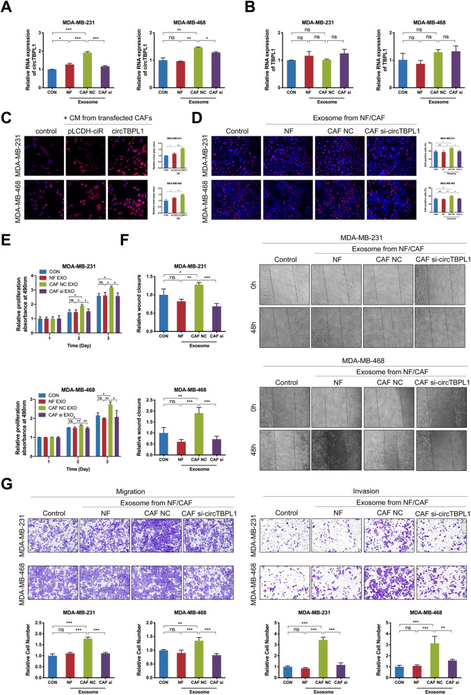
Breast cancer is the major common malignancy worldwide among women. Previous studies reported that cancer-associated fibroblasts (CAFs) showed pivotal roles in regulating tumor progression via exosome-mediated cellular communication. However, the detailed mechanism underlying the exosomal circRNA from CAFs in breast cancer progression remains ambiguous. Here, exosomal circRNA profiling of breast cancer-derived CAFs and normal fibroblasts (NFs) was detected by high-throughput sequencing, and upregulated circTBPL1 expression was identified in CAF exosomes. The exosomal circTBPL1 from CAFs could be transferred to breast cancer cells and promoted cell proliferation, migration, and invasion. Consistently, circTBPL1 knockdown in CAFs attenuated their tumor-promoting ability. Further exploration identified miR-653-5p as an inhibitory target of circTBPL1, and ectopic expression of miR-653-5p could partially reverse the malignant phenotypes induced by circTBPL1 overexpression in breast cancer. Additionally, TPBG was selected as a downstream target gene, and circTBPL1 could protect TPBG from miR-653-5p-mediated degradation, leading to enhanced breast cancer progression. Significantly, the accelerated tumor progression triggered by exosomal circTBPL1 from CAFs was confirmed in xenograft models. Taken together, these results revealed that exosomal circTBPL1 derived from CAFs contributed to cancer progression via miR-653-5p/TPBG pathway, indicating the potential of exosomal circTBPL1 as a biomarker and novel therapeutic target for breast cancer.


Conflict of interest statement

The authors declare no competing interests.

Figures

Fig. 1
Fig. 1. Exosomes derived from CAFs exhibit upregulated circTBPL1 expression.
A The Circos plot shows the differentially expressed circRNAs in exosomes derived from CAFs and NFs. The outermost circle shows the chromosomal distribution of the circRNAs. The second circle indicates the circBase ID of the predominantly enriched circRNAs in exosomes derived from CAFs (orange) or NFs (blue). The third circle shows the log2FC of differentially expressed circRNAs (orange squares for upregulated circRNAs and purple squares for downregulated circRNAs). The fourth circle shows the number of reads the differentially expressed circRNAs matched after normalization (orange circles for CAFs and blue triangles for NFs). B The qRT-PCR analysis showing the circTBPL1 expression in exosomes secreted by NFs and CAFs (left) and that in the corresponding NFs and CAFs (right). C ISH assay was performed to detect the localization of circTBPL1 in breast cancer tissue, and HE staining was used to show the histology. D Schematic illustration showing the genomic location and splicing pattern of circTBPL1. The back-splice junction (red triangle) of circTBPL1was confirmed by sanger sequencing. E cDNA and gDNA of CAFs were amplified using convergent and divergent primers. β-actin was used as the negative control. F The qRT–PCR analysis was used to detect the expression of circTBPL1 and linear TBPL1 in CAFs treated with actinomycin D for indicated time. G The qRT–PCR analysis was performed to evaluate the expression of circTBPL1 and linear TBPL1 in CAFs treated with RNase R. H The expression of circTBPL1 and linear TBPL1 were evaluated by qRT-PCR using random primers or oligo dT primers. I The expression of circTBPL1 in the cytoplasm and nucleus of CAFs was evaluated by qRT–PCR. GAPDH and U6 were used as cytoplasmic and nuclear RNA markers, respectively. J FISH assay was used to detect the subcellular localization of circTBPL1 in CAFs. (ns, no significance, *P < 0.05, **P < 0.01).
Fig. 2
Fig. 2. Exosomal circTBPL1 is essential for proliferation and metastasis of breast cancer cells.
A, B The qRT-PCR analysis of circTBPL1 (A) and linear TBPL1 (B) expression in breast cancer cells treated with or without indicated exosomes. C The expression level of circTBPL1 in breast cancer cells treated with CM from CAFs was detected by FISH assay. D, E Edu (D) and MTT (E) assays showed the effect of exosomes on the proliferation of breast cancer cell. F Wound healing assay indicated the influence of exosomes on the migration of breast cancer cells. G Transwell assay showed the impact of exosomes on breast cancer cell migration and invasion. (ns, no significance, *P < 0.05, **P < 0.01, ***P < 0.001).
Fig. 3
Fig. 3. circTBPL1 overexpression promotes cell proliferation and motility of breast cancer cells.
A The circTBPL1 overexpression efficiency and TBPL1 expression level were verified using qRT-PCR assay. BD The proliferation of breast cancer cells after circTBPL1 overexpression was detected by MTT assay (B), colony formation assay (C), and Edu assay (D). E Transwell assay was performed to evaluate the effect of circTBPL1 overexpression on the migration and invasion abilities of breast cancer cells. F Wound healing assay was used to detect the cell migration after circTBPL1 overexpression in breast cancer cells. G Phalloidin staining was used to show the morphological change of breast cancer cells transfected with pLCDH-ciR or circTBPL1. H The expression of EMT-related markers was assessed by western blot. I Tube formation assay was performed to evaluate the angiogenesis-promoting effect of circTBPL1 overexpression in breast cancer cells. (ns, no significance, *P < 0.05, **P < 0.01, ***P < 0.001).
Fig. 4
Fig. 4. circTBPL1 serves as a miRNA sponge for miR-653-5p in breast cancer cells.
A The expression of circTBPL1 in the cytoplasm and nucleus of breast cancer cells was evaluated by qRT–PCR. GAPDH and U6 were used as cytoplasmic and nuclear RNA markers, respectively. B FISH assay was used to detect the subcellular localization of circTBPL1 in breast cancer cells. C Venn diagram showing the potential circTBPL1-binding miRNAs, predicted by CircInteractome and Starbase databases. D The schematic diagram of luciferase reporter (top). The predicted circTBPL1-binding site with miR-653-5p and the corresponding mutant sequence (bottom). E The binding relationship between circTBPL1 and miR-653-5p was verified using the dual-luciferase reporter assay. F RIP assay detected the enrichment of circTBPL1 and miR-653-5p in breast cancer cells. G RNA-FISH showed that circTBPL1 was colocalized with miR-653-5p in breast cancer cells. H The miR-653-5p expression after circTBPL1 knockdown in breast cancer cells. (ns, no significance, *P < 0.05, **P < 0.01, ***P < 0.001).
Fig. 5
Fig. 5. circTBPL1 promotes breast cancer progression through regulating miR-653-5p.
A The transfection efficiency of circTBPL1 and miR-653-5p in breast cancer cells was detected using qRT-PCR. BD MTT assay (B), colony formation assay (C), and Edu assay (D) were used to evaluate the proliferation ability of breast cancer cells in different transfected group. E Wound healing assay showed the migration and invasion abilities of breast cancer cells. F Transwell experiment was used to detect the metastasis ability of breast cancer cells. G Tube formation assay was performed to evaluate the angiogenesis-promoting effect of breast cancer cells. (ns, no significance, *P < 0.05, **P < 0.01, ***P < 0.001).
Fig. 6
Fig. 6. circTBPL1 sequesters miR-653-5p to promote TPBG expression.
A Venn diagram showing the potential downstream target genes of miR-653-5p, predicted by DIANA, miRDB, mirDIP, and Starbase databases (top). The predicted miR-653-5p-binding sites with TPBG and the corresponding mutant sequence (bottom). B, C The mRNA and protein levels of TPBG after circTBPL1 overexpression (B) or circTBPL1 knockdown (C) were detected. D The mRNA and protein levels of TPBG after miR-653-5p overexpression were evaluated. E Following circTBPL1 overexpression and miR-653-5p mimics transfection, the mRNA and protein expression of TPBG was measured. F The binding relationship between miR-653-5p and TPBG was verified using the dual-luciferase reporter assay. G The overexpression efficiency of TPBG was evaluated using qRT-PCR and western blot. HJ Cellular proliferation following TPBG overexpression was assessed via MTT assay (H), colony formation assay (I), and Edu assay (J). K Wound healing assay was employed to appraise cell migration. L Transwell assay was implemented to analyze cell migration and invasion. M The expression of EMT-related markers in these cells was examined via western blot. (ns, no significance, *P < 0.05, **P < 0.01, ***P < 0.001).
Fig. 7
Fig. 7. circTBPL1 promotes malignant behaviors of breast cancer cells via regulating TPBG expression.
Breast cancer cells were co-transfected with circTBPL1 overexpression vectors and TPBG siRNA. A The RNA and protein expression of TPBG was detected using qRT-PCR and western blot. BD The proliferation ability of breast cancer cells was evaluated by MTT assay (B), colony formation assay (C), and Edu assay (D). E Wound healing assay was performed to investigate the migration ability of breast cancer cells. F Transwell assay was used to evaluate the migration and invasion abilities of breast cancer cells. (ns, no significance, *P < 0.05, **P < 0.01, ***P < 0.001).
Fig. 8
Fig. 8. Exosomal circTBPL1 derived from CAFs regulates miR-653-5p/TPBG axis in vivo to promote tumor growth and metastasis of breast cancer.
A Nude mice were subcutaneously implanted with MDA-MB-231 cells co-injected with CAF pLCDH-ciR or CAF circTBPL1. The images of xenograft tumors in each group. B, C Tumor volume (B) and weight (C) were measured in each group. D, E qRT-PCR (D) and western blot (E) were used to detect the expression of related factors in tumor tissues. F Tumor tissues from xenograft model mice were subjected to HE staining, and IHC staining was performed to evaluate the expression of the related factors in tumor tissues. G Representative images of the lung tissues after tail vein injection of MDA-MB-231 cells with or without circTBPL1 overexpression. H Representative images of HE staining for lung tissues in each group. I The histogram analysis of the metastatic nodules number in per lung. J The schematic overview illustrates the mechanistic basis for the observational study results. CAF-derived circTBPL1 is transmitted into breast cancer cells through exosomes and can therein modulate the miR-653-5p/TPBG axis to influence tumor growth and metastasis. (**P < 0.01, ***P < 0.001).

Similar articles

Cited by

References

    1. Siegel RL, Miller KD, Fuchs HE, Jemal A. Cancer statistics, 2022. CA Cancer J Clin. 2022;72:7–33. doi: 10.3322/caac.21708. - DOI - PubMed
    1. Zhang N, Cao C, Zhu Y, Liu P, Liu L, Lu K, et al. Primary breast lymphoma: a single center study. Oncol Lett. 2017;13:1014–8. doi: 10.3892/ol.2016.5483. - DOI - PMC - PubMed
    1. Harbeck N, Penault-Llorca F, Cortes J, Gnant M, Houssami N, Poortmans P, et al. Breast cancer. Nat Rev Dis Prim. 2019;5:66. doi: 10.1038/s41572-019-0111-2. - DOI - PubMed
    1. Valastyan S, Weinberg RA. Tumor metastasis: molecular insights and evolving paradigms. Cell. 2011;147:275–92. doi: 10.1016/j.cell.2011.09.024. - DOI - PMC - PubMed
    1. Mittal S, Brown NJ, Holen I. The breast tumor microenvironment: role in cancer development, progression and response to therapy. Expert Rev Mol Diagn. 2018;18:227–43. doi: 10.1080/14737159.2018.1439382. - DOI - PubMed

Publication types