New microvascular ultrasound techniques: abdominal applications
- PMID: 37495910
- PMCID: PMC10473992
- DOI: 10.1007/s11547-023-01679-6
New microvascular ultrasound techniques: abdominal applications
Abstract
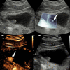
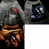
Microvascular ultrasound (MVUS) is a new ultrasound technique that allows the detection of slow-velocity flow, providing the visualization of the blood flow in small vessels without the need of intravenous contrast agent administration. This technology has been integrated in the most recent ultrasound equipment and applied for the assessment of vascularization. Compared to conventional color Doppler and power Doppler imaging, MVUS provides higher capability to detect intralesional flow. A growing number of studies explored the potential applications in hepatobiliary, genitourinary, and vascular pathologies. Different flow patterns can be observed in hepatic and renal focal lesions providing information on tumor vascularity and improving the differential diagnosis. This article aims to provide a detailed review on the current evidences and applications of MVUS in abdominal imaging.
Keywords: Contrast-enhanced ultrasound; Micro-flow imaging; Microvascular flow imaging; Superb microvascular imaging.
© 2023. The Author(s).
Conflict of interest statement
Roberto Cannella has the following disclosures: support for attending meetings from Bracco and Bayer; research collaboration with Siemens Healthcare; co-funding by the European Union—FESR or FSE, PON Research and Innovation 2014–2020—DM 1062/2021.
Figures
Similar articles
-
Evaluation of Ovarian Vascularity in Children by Using the "Superb Microvascular Imaging" Ultrasound Technique in Comparison With Conventional Doppler Ultrasound Techniques.J Ultrasound Med. 2019 Oct;38(10):2751-2760. doi: 10.1002/jum.14983. Epub 2019 Mar 28. J Ultrasound Med. 2019. PMID: 30919993
-
Comparison of Superb Microvascular Imaging With Other Doppler Methods in Assessment of Testicular Vascularity in Cryptorchidism.Ultrasound Q. 2020 Dec;36(4):363-370. doi: 10.1097/RUQ.0000000000000533. Ultrasound Q. 2020. PMID: 32956243
-
Comparison of Superb Microvascular Imaging With Color Flow and Power Doppler Imaging of Small Hepatocellular Carcinomas.J Ultrasound Med. 2018 Dec;37(12):2915-2924. doi: 10.1002/jum.14654. Epub 2018 Apr 23. J Ultrasound Med. 2018. PMID: 29683199
-
Ultrasonographic Demonstration of the Tissue Microvasculature in Children: Microvascular Ultrasonography Versus Conventional Color Doppler Ultrasonography.Korean J Radiol. 2020 Feb;21(2):146-158. doi: 10.3348/kjr.2019.0500. Korean J Radiol. 2020. PMID: 31997590 Free PMC article. Review.
-
The clinical application of ultrasonography with superb microvascular imaging-a review.J Clin Ultrasound. 2022 Jun;50(5):721-732. doi: 10.1002/jcu.23210. Epub 2022 Mar 31. J Clin Ultrasound. 2022. PMID: 35358353 Review.
Cited by
-
Diagnostic accuracy of microvascular flow imaging ultrasound for endoleak detection after endovascular aortic aneurysm repair: a systematic review and meta-analysis.Pol J Radiol. 2024 Aug 22;89:e414-e419. doi: 10.5114/pjr/190502. eCollection 2024. Pol J Radiol. 2024. PMID: 39257925 Free PMC article. Review.
-
The global burden of vascular intestinal diseases: results from the 2021 Global Burden of Disease Study and projections using Bayesian age-period-cohort analysis.Environ Health Prev Med. 2024;29:71. doi: 10.1265/ehpm.24-00206. Environ Health Prev Med. 2024. PMID: 39662956 Free PMC article.
-
Breast multiparametric ultrasound: a single-center experience.J Ultrasound. 2024 Dec;27(4):831-839. doi: 10.1007/s40477-024-00944-2. Epub 2024 Aug 5. J Ultrasound. 2024. PMID: 39103741
-
Diagnostic performance of microvascular flow imaging for noninvasive assessment of liver fibrosis in chronic liver disease.PLoS One. 2025 Jun 4;20(6):e0322102. doi: 10.1371/journal.pone.0322102. eCollection 2025. PLoS One. 2025. PMID: 40465598 Free PMC article.
-
Radiological Assessment After Pancreaticoduodenectomy for a Precision Approach to Managing Complications: A Narrative Review.J Pers Med. 2025 May 28;15(6):220. doi: 10.3390/jpm15060220. J Pers Med. 2025. PMID: 40559083 Free PMC article. Review.
References
Publication types
MeSH terms
LinkOut - more resources
Full Text Sources
Medical

