. 2023 Jul 5;5(15):3914-3923.
doi: 10.1039/d3na00124e. eCollection 2023 Jul 25.

Charge controlled interactions between DNA-modified silica nanoparticles and fluorosurfactants in microfluidic water-in-oil droplets

Affiliations

Charge controlled interactions between DNA-modified silica nanoparticles and fluorosurfactants in microfluidic water-in-oil droplets

Sahana Sheshachala et al. Nanoscale Adv. .

Abstract

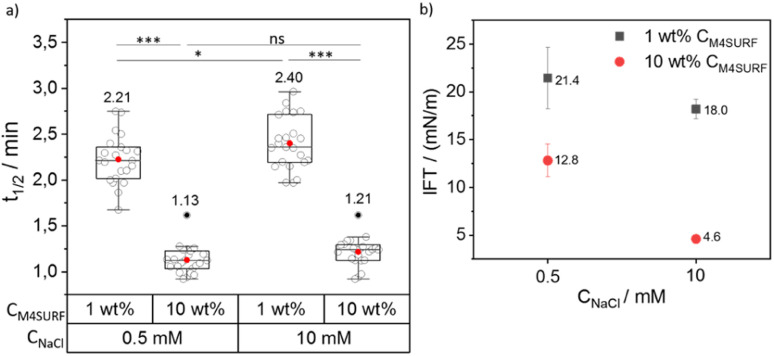
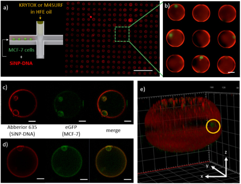
Microfluidic droplets are an important tool for studying and mimicking biological systems, e.g., to examine with high throughput the interaction of biomolecular components and the functionality of natural cells, or to develop basic principles for the engineering of artificial cells. Of particular importance is the approach to generate a biomimetic membrane by supramolecular self-assembly of nanoparticle components dissolved in the aqueous phase of the droplets at the inner water/oil interface, which can serve both to mechanically reinforce the droplets and as an interaction surface for cells and other components. While this interfacial assembly driven by electrostatic interaction of surfactants is quite well developed for water/mineral oil (W/MO) systems, no approaches have yet been described to exploit this principle for water/fluorocarbon oil (W/FO) emulsion droplets. Since W/FO systems exhibit not only better compartmentalization but also gas solubility properties, which is particularly crucial for live cell encapsulation and cultivation, we report here the investigation of charged fluorosurfactants for the self-assembly of DNA-modified silica nanoparticles (SiNP-DNA) at the interface of microfluidic W/FO emulsions. To this end, an efficient multicomponent Ugi reaction was used to synthesize the novel fluorosurfactant M4SURF to study the segregation and accumulation of negatively charged SiNP-DNA at the inner interface of microfluidic droplets. Comparative measurements were performed with the negatively charged fluorosurfactant KRYTOX, which can also induce SiNP-DNA segregation in the presence of cations. The segregation dynamics is characterized and preliminary results of cell encapsulation in the SiNP-DNA functionalized droplets are shown.


Conflict of interest statement

There are no conflicts to declare.

Figures

Fig. 1
Fig. 1. (a) Chemical structures of positively charged M4SURF (1), the neutral PFPE–PEG–PFPE type fluorosurfactant PICO-SURF (2) and negatively charged KRYTOX (3). (b) Schematic illustration of the 4-component Ugi reaction to synthesize M4SURF. Details of the synthesis can be found in the Experimental section and in Fig. S1 in the ESI, which also includes a discussion on the efficiency and sustainability of the multicomponent Ugi reaction.
Fig. 2
Fig. 2. (a) Schematics of the charge-induced segregation of colloidal silica-DNA nanoparticles (SiNP-DNA) dispersed in PBS buffer containing 10 mM NaCl; (b–e) fluorescence images of W/FO droplets in the presence of three different surfactants, PICO-SURF (b and d), KRYTOX (c) and M4SURF (e). Note that the droplets in (c) and (e) were formed from the droplets in (b) and (d) after in situ addition of KRYTOX and M4SURF, respectively. Scale bars are 100 μm.
Fig. 3
Fig. 3. (a) Grouped box plot of t1/2 values obtained for the SiNP-DNA interface assembly with a varying weight percentage (1 wt% and 10 wt%) of KRYTOX in HFE 7500 oil against concentrations of 0.5 and 10 mM NaCl. The box plot shows the median and 25–75th percentiles. Individual data points, mean and outliers are represented by hollow, red and black dots, respectively. 33 droplets were analyzed from 5 independent experiments for each condition. The corresponding mean values are also indicated above each box plot. (b) Interfacial tension (IFT) values for SiNP-DNA/KRYTOX systems obtained at the variable concentrations of KRYTOX and NaCl used in this study, as analyzed using 10–15 pendant droplets. A two-way ANOVA was performed using SPSS software to analyze the effect of CNaCl and CKRYTOX on the t1/2. The test revealed that there was no statistically significant interaction between the effects of CNaCl and CKRYTOX (p = 0.181). Simple main effects analysis showed significant effects on the t1/2 that are indicated by asterisks (***p < 0.001). Outliers were determined according to Tukey's formula [Q1 − 1.5 IQR; Q3 + 1.5 IQR].
Fig. 4
Fig. 4. (a) Grouped box plot of t1/2 values obtained for SiNP-DNA interface assembly with a varying weight percentage (1 wt% and 10 wt%) of M4SURF (a) in HFE 7500 oil, against concentrations of 0.5 and 10 mM NaCl. The box plot shows the median and 25–75th percentiles. Individual data points, mean and outliers are represented by hollow, red and black dots, respectively. 23 droplets were analyzed from 4 independent experiments for each condition. The corresponding mean values are also indicated above each box plot. (b) Interfacial tension (IFT) values for SiNP-DNA/M4SURF systems obtained at the variable concentrations of M4SURF and NaCl used in this study, as analyzed using 10–15 pendant droplets. A two-way ANOVA was performed using SPSS software to analyze the effect of CNaCl and CM4SURF on the t1/2. The test revealed that there was no statistically significant interaction between the effects of CNaCl and CM4SURF (p = 0.356). Simple main effects analysis showed significant effects on the t1/2 that are indicated by asterisks (*p < 0.05; ***p < 0.001). Outliers were determined according to Tukey's formula [Q1 – 1.5 IQR; Q3 + 1.5 IQR].
Fig. 5
Fig. 5. (a) Schematic representation of the encapsulation of MCF-7eGFP cells with SiNP-DNA in cell culture medium (left) and a representative fluorescence image of the microfluidic chip containing an array of droplets inside the on-chip storage wells (right). The scale bar is 500 μm. (b) Enlarged image of microfluidic droplets containing MCF-7 cells and self-assembled SiNP-DNA. Scale bar is 50 μm. (c and d) 2D confocal images of the MCF-7eGFP cells (green) inside microfluidic droplets obtained with the SiNP-DNA/KRYTOX (c) and SiNP/M4SURF (d) systems. The shell-like structures (red) contain the MCF-7eGFP cells (green) that adhered to the SiNP-DNA-decorated interface. Scale bars are 25 μm. (e) 3D reconstruction image of a representative microfluidic droplet containing an encapsulated MCF-7eGFP cell (green, marked by a yellow circle) and the self-assembled SiNP-DNA layer.

Similar articles

Cited by

  • Control of Buckling of Colloidal Supraparticles.
    Roemling LJ, De Angelis G, Mauch A, Amstad E, Vogel N. Roemling LJ, et al. Small. 2025 Jun;21(22):e2411772. doi: 10.1002/smll.202411772. Epub 2025 May 2. Small. 2025. PMID: 40317860 Free PMC article.

References

    1. Sackmann E. K. Fulton A. L. Beebe D. J. Nature. 2014;507:181–189. doi: 10.1038/nature13118. - DOI - PubMed
    1. Chou W.-L. Lee P.-Y. Yang C.-L. Huang W.-Y. Lin Y.-S. Micromachines. 2015;6:1249. doi: 10.3390/mi6091249. - DOI
    1. Martino C. deMello A. J. Interface Focus. 2016;6:20160011. doi: 10.1098/rsfs.2016.0011. - DOI - PMC - PubMed
    1. Göpfrich K. Platzman I. Spatz J. P. Trends Biotechnol. 2018;36:938–951. doi: 10.1016/j.tibtech.2018.03.008. - DOI - PMC - PubMed
    1. Dinsmore A. D. Ming F. H. Nikolaides M. G. Marquez M. Bausch A. R. Weitz D. A. Science. 2002;298:1006–1009. doi: 10.1126/science.1074868. - DOI - PubMed