. 2023 Jun 20;5(15):3896-3904.
doi: 10.1039/d3na00251a. eCollection 2023 Jul 25.

The luminescence mechanism of ligand-induced interface states in silicon quantum dots

Affiliations

The luminescence mechanism of ligand-induced interface states in silicon quantum dots

Jian Zhou et al. Nanoscale Adv. .

Abstract

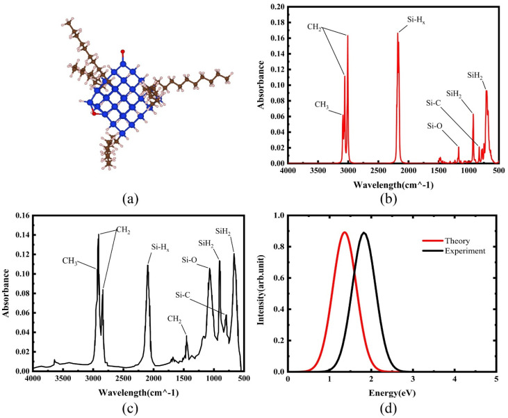
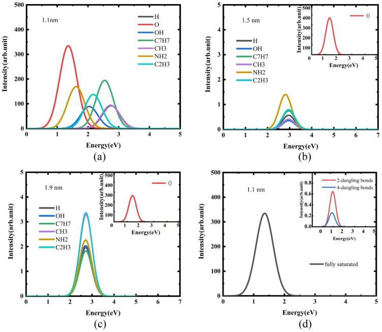
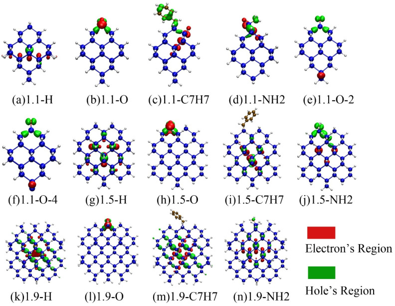
Over decades of research on photoluminescence (PL) of silicon quantum dots (Si-QDs), extensive exploratory experiments have been conducted to find ways to improve the photoluminescence quantum yield. However, the complete physical picture of Si-QD luminescence is not yet clear and needs to be studied in depth. In this work, which considers the quantum size effect and surface effect, the optical properties of Si-QDs with different sizes and surface terminated ligands were calculated based on first principles calculations. The results show that there are significant differences in the emission wavelength and emission intensity of Si-QD interface states connected by different ligands, among which the emission of silicon-oxygen double bonds is the strongest. When the size of the Si-QD increases, the influence of the surface effect weakens, and only the silicon-oxygen double bonds still localize the charge near the ligand, maintaining a high-intensity luminescence. In addition, the presence of surface dangling bonds also affects luminescence. This study deepens the understanding of the photoluminescence mechanism of Si-QDs, and provides a direction for both future improvement of the photoluminescence quantum efficiency of silicon nanocrystals and for fabricating silicon-based photonic devices.


Conflict of interest statement

There are no conflicts to declare.

Figures

Fig. 1
Fig. 1. Schematic diagram of the structure of Si-QDs terminated by hydrogens and one oxygen double bond of different sizes: (a) 1.1 nm diameter, (b) 1.5 nm diameter, and (c) 1.9 nm diameter.
Fig. 2
Fig. 2. (a) Structural model and (b) FTIR spectrum of Si-QDs by theoretical calculations and (c) FTIR spectrum of Si-QDs measured by experiment. Reproduced with permission from ref. , copyright American Chemical Society, 2022. (d) PL spectra of Si-QDs obtained theoretically and experimentally, where the black line is the experimental result in ref. , and the red line is the result of theoretical calculations (peak height is normalized).
Fig. 3
Fig. 3. PL spectra of quantum dots of different sizes when their surfaces are terminated by different ligands: (a) 1.1 nm diameter, (b) 1.5 nm diameter, (c) 1.9 nm diameter, and (d) 1.1 nm diameter quantum dots with different numbers of dangling bonds terminated by silicon–oxygen double bonds.
Fig. 4
Fig. 4. (a)–(d) and (g)–(n) Schematic diagram of the electron–hole distribution in the emission spectrum of quantum dots with different sizes and different ligand terminations. (e) and (f) Quantum dots with dangling bonds on the surface, with the remainder saturated on the surface. Red represents the electron distribution area, and green represents the hole distribution area.
Fig. 5
Fig. 5. Projection of molecular orbitals of silicon quantum dots of different sizes, terminated by different surface ligands in the ΓX direction of the Brillouin zone. (a)–(c) Silicon quantum dots with surfaces of 1.1, 1.5 and 1.9 nm terminated only by hydrogen. (d)–(f) 1.1, 1.5 and 1.9 nm silicon quantum dots terminated by silicon–oxygen double bonds on the surface. The color bar is used to represent the density of the corresponding state; the darker the color, the more electronic states there are.

References

    1. Hawkins A. R. Reynolds T. E. England D. R. Babic D. I. Mondry M. J. Streubel K. Bowers J. E. Appl. Phys. Lett. 1996;68:3692–3694. doi: 10.1063/1.115975. - DOI
    1. Xin Y. Nishio K. Saitow K.-I. Appl. Phys. Lett. 2015;106:201102. doi: 10.1063/1.4921415. - DOI
    1. Wang D. Zhang C. Zeng P. Zhou W. Ma L. Wang H. Zhou Z.-q. Hu F. Zhang S. Lu M. Wu X. J. C. S. B. Sci. Bull. 2018;63:75–77. doi: 10.1016/j.scib.2018.01.006. - DOI - PubMed
    1. Zhang C. Yang B. Chen J. Wang D. Zhang Y. Li S. Dai X. Zhang S. Lu M. Opt. Express. 2020;28:194–204. doi: 10.1364/OE.382691. - DOI - PubMed
    1. Ono T. Xu Y. Sakata T. Saitow K.-I. ACS Appl. Mater. Interfaces. 2021;14:1373–1388. doi: 10.1021/acsami.1c18779. - DOI - PubMed