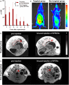
Endometriosis-targeted MRI imaging using bevacizumab-modified nanoparticles aimed at vascular endothelial growth factor
- PMID: 37496625
- PMCID: PMC10367955
- DOI: 10.1039/d2na00787h
Endometriosis-targeted MRI imaging using bevacizumab-modified nanoparticles aimed at vascular endothelial growth factor
Abstract
Endometriosis is a tumor-like disease with high recurrence. In this case, the accurate imaging-based diagnosis of endometriosis can help clinicians eradicate it by improving their surgical plan. However, although contrast agents can improve the visibility of the tissue of interest in vivo via magnetic resonance imaging (MRI), the lack of biomarkers in endometriosis hinders the development of agents for its targeted imaging and diagnosis. Herein, aiming at the enriched vascular endothelial growth factor (VEGF) in endometriosis, we developed a targeting MRI contrast agent modified with bevacizumab, i.e., NaGdF4@PEG@bevacizumab-Cy5.5 nanoparticles (NPBCNs), to detect endometriosis. NPBCNs showed negligible cytotoxicity and high affinity towards VEGF in endometrial cells in vitro. Furthermore, NPBCNs generated a strong signal enhancement in vivo in endometriosis lesions in rats in T1-weighted images via MRI at 3 days post-injection, as confirmed by the histopathological staining results and fluorescence imaging on the same day. Our approach can enable NPBCNs to target endometriosis effectively, thus avoiding missed diagnoses.
This journal is © The Royal Society of Chemistry.
Conflict of interest statement
The authors have declared that no conflict of interest exists.
Figures


Similar articles
-
Effect of vascular endothelial growth factor inhibition on endometrial implant development in a murine model of endometriosis.Reprod Sci. 2011 Jul;18(7):614-22. doi: 10.1177/1933719110395406. Epub 2011 Jan 25. Reprod Sci. 2011. PMID: 21266664
-
Targeted Nanoparticles with High Heating Efficiency for the Treatment of Endometriosis with Systemically Delivered Magnetic Hyperthermia.Small. 2022 Jun;18(24):e2107808. doi: 10.1002/smll.202107808. Epub 2022 Apr 17. Small. 2022. PMID: 35434932 Free PMC article.
-
Bevacizumab and near infrared probe conjugated iron oxide nanoparticles for vascular endothelial growth factor targeted MR and optical imaging.Biomater Sci. 2018 May 29;6(6):1517-1525. doi: 10.1039/c8bm00225h. Biomater Sci. 2018. PMID: 29652061 Free PMC article.
-
Magnetic resonance imaging characteristics of polypoid endometriosis and review of the literature.J Obstet Gynaecol Res. 2022 Oct;48(10):2583-2593. doi: 10.1111/jog.15367. Epub 2022 Jul 22. J Obstet Gynaecol Res. 2022. PMID: 35868869 Review.
-
Monoclonal antibodies targeting vascular endothelial growth factor: current status and future challenges in cancer therapy.BioDrugs. 2009;23(5):289-304. doi: 10.2165/11317600-000000000-00000. BioDrugs. 2009. PMID: 19754219 Review.
Cited by
-
The Role of Nanomedicine in Benign Gynecologic Disorders.Molecules. 2024 May 1;29(9):2095. doi: 10.3390/molecules29092095. Molecules. 2024. PMID: 38731586 Free PMC article. Review.
References
-
- David Adamson G. Kennedy S. Hummelshoj L. J. Endometr. 2010;2:3–6. doi: 10.1177/228402651000200102. - DOI
LinkOut - more resources
Full Text Sources

