. 2023 Jun 28;26(7):107236.
doi: 10.1016/j.isci.2023.107236. eCollection 2023 Jul 21.

Human neutrophils communicate remotely via calcium-dependent glutamate-induced glutamate release

Affiliations

Human neutrophils communicate remotely via calcium-dependent glutamate-induced glutamate release

Olga Kopach et al. iScience. .

Abstract

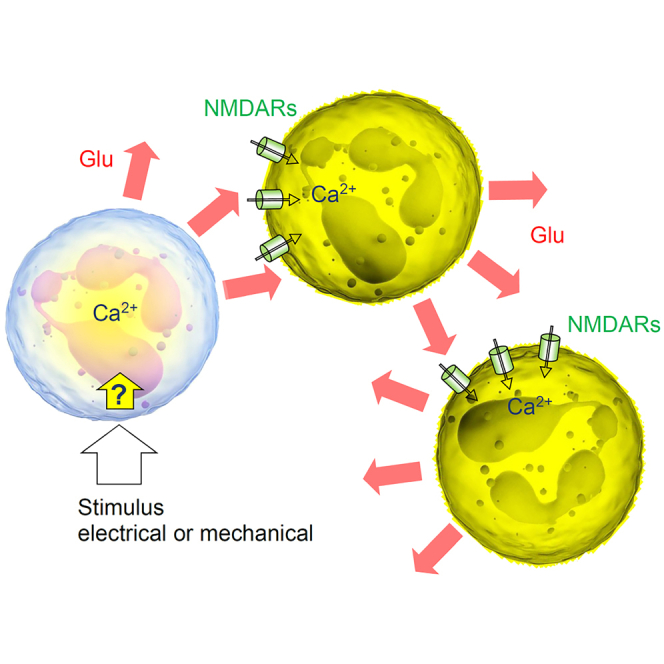
Neutrophils are white blood cells that are critical to acute inflammatory and adaptive immune responses. Their swarming-pattern behavior is controlled by multiple cellular cascades involving calcium-dependent release of various signaling molecules. Previous studies have reported that neutrophils express glutamate receptors and can release glutamate but evidence of direct neutrophil-neutrophil communication has been elusive. Here, we hold semi-suspended cultured human neutrophils in patch-clamp whole-cell mode to find that calcium mobilization induced by stimulating one neutrophil can trigger an N-methyl-D-aspartate (NMDA) receptor-driven membrane current and calcium signal in neighboring neutrophils. We employ an enzymatic-based imaging assay to image, in real time, glutamate release from neutrophils induced by glutamate released from their neighbors. These observations provide direct evidence for a positive-feedback inter-neutrophil communication that could contribute to mechanisms regulating communal neutrophil behavior.

Keywords: biological sciences; cell biology; immunology; molecular biology; neuroscience.


Conflict of interest statement

The authors declare no competing interests.

Figures

None
Graphical abstract
Figure 1
Figure 1
NMDA receptor-mediated Ca2+ entry in human neutrophils (A) Diagram depicting probing of NMDARs with a rapid-exchange system [28]: a neutrophil (held in whole-cell) is stimulated pharmacologically by applying different solutions through two channels of ϴ-glass pipette (tip diameter ∼200 μm) mounted on a piezo-drive to enable the ultra-fast delivery (<1 ms resolution; solutions in ϴ glass exchanged within 10 s using a rapid multi-channel perfusion system); see Videos S1 and S2 for live videos of neutrophils before and during patching. (B) Representative whole-cell currents (patch pipette 4–7 MΩ, 1–2 μM tip) recorded in human neutrophils in response to locally applied NMDA (1 mM) and glycine (1 mM), in zero Mg2+, 2 mM Mg2+, and in the presence of NMDAR antagonists APV (50 μM) and Co101244 (1 μM) in zero Mg2+ (control), as indicated; same-cell pharmacological manipulations applied at ∼20 s intervals. (C) Summary of experiments shown in (B); mean ± SEM (amplitude over the 300–500 ms pulse segment), normalized to control (sample size shown); ∗∗∗p < 0.01. Inset, super-resolution dSTORM image of a neutrophil shown with chromatically separated GluN2B and elastase single-molecule labels as indicated; see Figure S1A for further detail and illustrations. (D) Characteristic images of a neutrophil (gray DIC image, top raw) loaded with CellTracker Red (red channel, middle) and Fluo 4-AM (green, bottom), in baseline conditions, after bath application of NMDA (100 μM, 2-3-min duration) and glycine (50 μM), and after adding PMA (1 μM) for Ca2+ homeostasis control, as indicated; experimental timing as shown; false color scale: relative intensity, arbitrary units (au); pixel size ∼120 nm (near diffraction limit). (E) Experiment as in (D), but in the presence of the selective GluN2B-containing NMDAR antagonist Co101244 (1 μM); other notations as in (D). (F) Statistical summary of experiments shown in (D and E). Average Ca2+ responses (mean ± SEM) of individual neutrophils, first to NMDA+Gly application (blue) and next to PMA (red) application, in either of the three solutions: 0 Mg2+ (as in D), 2 mM Mg2+, and 0 Mg2+ with Co101244 (as in E), as indicated; no NMDA+Gly was applied to control sample (Cntrl, green; 0 Mg2+), to evaluate an experiment-wise drift in Ca2+-dependent fluorescence at the time of NMDA+Gly response measurement in other groups (dotted line); numbers, sample size (in 0 Mg2+ group, 3 cells were lost at the PMA stage); ∗∗∗p < 0.001 (paired t test; two-sample t test for 0 Mg2+ group). (G) One-cell example (neutrophil held in whole-cell, patch pipette is seen) showing that depolarization current induces prominent Ca2+ mobilization; inset, DIC+Fluo-4 AM image; traces: F, Fluo-4 fluorescence signal; Ih, holding current. (H) Summary of experiments shown in (G). Left: Snapshots (Fluo-4 channel) of the stimulated cell (red dot; a neighboring neutrophil can be seen), in control conditions (Cntrl) and in the presence of APV, as indicated, at the time points as indicated (seconds) with respect to the stimulus onset. Note that in control conditions, but not under APV, the neighboring cell responds with a Ca2+ rise. Right: Summary of depolarization-induced [Ca2+]in rises in the patched cell, represented by the Fluo-4 ΔF/F0 signal; dots, individual cells; bars: mean ± SEM; sample size shown.
Figure 2
Figure 2
Stimulation of one neutrophil elicits NMDAR-mediated currents, Ca2+ rise, and glutamate release in neighboring neutrophils (A) Illustration, dual whole-cell recordings from two human neutrophils (cell 1 and cell 2) semi-suspended in culture; patch-pipette tips can be seen; some neutrophils lie destroyed after unsuccessful dual-patch attempts. (B) Example traces from experiments in (A). A 50 ms current pulse applied in cell 1 (upper trace; current clamp) triggers an inward current deflection in cell 2 (voltage clamp), which is blocked by 50 μM APV; bath medium contains 1 mM glycine. (C) Summary of experiments shown in (A and B) for the five recorded neutrophil pairs (left), and theoretical estimates of the glutamate concentration time course at three different distances, as indicated, from a small source neutrophil (right); see supplemental methods for detail. (D) Single-cell induced spread of Ca2+ signals among neutrophils. Time-lapse sequence (green Fluo-4 channel) in a semi-suspension of human neutrophils; dotted circle, neutrophil held in whole-cell mode (patch pipette fragment from DIC image shown) enabling one-cell electrical stimulation; electric stimulus (50 pA current) is applied at time zero to the patched cell; false color scale of Fluo-4 fluorescence F, as indicated; see Figure S1 for characteristic traces and Video S3 for the recorded image sequence. (E) Statistical summary of experiments in (D): amplitude of Fluo-4 fluorescence response (peak response; mean ± SEM) at the stimulated cell (Stim), neighboring intact neutrophils (Intact), and in the presence of the NMDAR blocker 3-((+)-2-carboxypiperazin-4-yl)propyl-l-phosphonic acid (CPP, 10 μM; +CPP) or the wide-range metabotropic glutamate receptor blocker alpha-methyl-4-carboxyphenylglycine (MCPG, 400 μM, + MCPG); numbers inside bars, sample size; ∗∗∗, p < 0.001 (two-sample t test, with respect to the three other sample means); see Videos S4 and S5 for recoded examples in CPP and MCPG samples. Note that peak responses were detected in different cells at different times post-stimulus. The data represent 26 independent tests (coverslips) from 10 individual blood cell preparations. (F) Left: Glutamate biosensor sensitivity test: a characteristic response to a 10 μM glutamate application, as indicated; dots, sensor record taken every 15 s. Right: A glutamate biosensor record during 10 s whole-cell current injection (within 1 min from the sensor recording onset; indicated by red cell-patch diagram) in an individual neutrophil held in whole-cell, as in (D), with sensor recording continuing for approximately 5 min (see Figures S2B–S2D for further detail). (G) Top: Schematic of the glutamate imaging method; A fluorescent enzymatic-based assay involves conversion of β-nicotinamide adenine dinucleotide (NAD) by L-glutamic dehydrogenase (GDH) (in the presence of glutamate) to NADH that fluoresces upon UV light excitation. Bottom: Assay sensitivity text; a fluorescence response to a pressure pulse of 5 μM glutamate (∼1 μm pipette tip; fluorescence intensity average over a 10 μm × 10 μm area around the tip, to roughly represent the vicinity of an individual neutrophil); see Figure S2 for fluorescent time-lapse snapshots. (H) Time-lapse series of fluorescence images (NADH channel) depicting a prominent rise in extracellular glutamate near the two neutrophils upon mechanical stimulation (light touch by a 1 μm micropipette tip) of one cell (dotted circle) at t = 0 s, as indicated; false color scale; note that the glutamate concentration signal drops sharply away from the cell surfaces reflecting rapid dilution in the bath medium. (I) Statistical summary of experiments in (H): amplitude of glutamate-sensitive NADH fluorescence response (mean ± SEM) at the stimulated cell (Stim), neighboring intact neutrophils (Intact), and in the presence of the NMDAR blocker CPP (10 μM; +CPP); numbers inside bars, sample size; ∗∗∗, p < 0.001 (two-sample t test). The data represent 17 independent tests (coverslips) from 3 individual blood cell preparations; see Videos S6 and S7 for the illustration of recordings in Intact and CPP samples.

Similar articles

References

    1. Amulic B., Cazalet C., Hayes G.L., Metzler K.D., Zychlinsky A. Neutrophil function: from mechanisms to disease. Annu. Rev. Immunol. 2012;30:459–489. - PubMed
    1. Kolaczkowska E., Kubes P. Neutrophil recruitment and function in health and inflammation. Nat. Rev. Immunol. 2013;13:159–175. - PubMed
    1. Ng L.G., Ostuni R., Hidalgo A. Heterogeneity of neutrophils. Nat. Rev. Immunol. 2019;19:255–265. - PubMed
    1. Häger M., Cowland J.B., Borregaard N. Neutrophil granules in health and disease. J. Intern. Med. 2010;268:25–34. - PubMed
    1. Phillipson M., Kubes P. The neutrophil in vascular inflammation. Nat. Med. 2011;17:1381–1390. - PMC - PubMed

LinkOut - more resources